


High resolution crystal structure of proteinase K with thiourea
Ahmad, M.S., Akbar, Z., Choudhary, M.I.(2019) Bioorganic Chemistry
Experimental Data Snapshot
(2019) Bioorganic Chemistry
Entity ID: 1 | |||||
|---|---|---|---|---|---|
| Molecule | Chains | Sequence Length | Organism | Details | Image |
| Proteinase K | A [auth E] | 279 | Parengyodontium album | Mutation(s): 1 Gene Names: PROK EC: 3.4.21.64 | 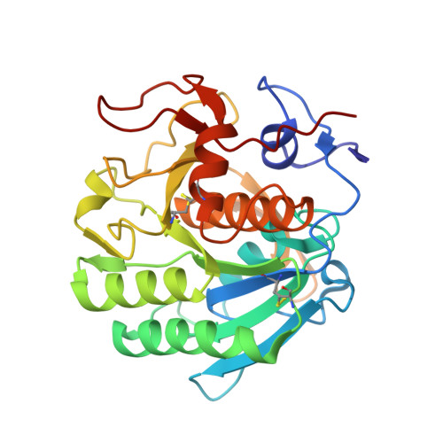 |
UniProt | |||||
Find proteins for P06873 (Parengyodontium album) Explore P06873 Go to UniProtKB: P06873 | |||||
Entity Groups | |||||
| Sequence Clusters | 30% Identity50% Identity70% Identity90% Identity95% Identity100% Identity | ||||
| UniProt Group | P06873 | ||||
Sequence AnnotationsExpand | |||||
| |||||
| Ligands 3 Unique | |||||
|---|---|---|---|---|---|
| ID | Chains | Name / Formula / InChI Key | 2D Diagram | 3D Interactions | |
| SO4 Query on SO4 | E | SULFATE ION O4 S QAOWNCQODCNURD-UHFFFAOYSA-L |  | ||
| TOU (Subject of Investigation/LOI) Query on TOU | B [auth E], C [auth E], D [auth E] | THIOUREA C H4 N2 S UMGDCJDMYOKAJW-UHFFFAOYSA-N |  | ||
| CL Query on CL | F [auth E] | CHLORIDE ION Cl VEXZGXHMUGYJMC-UHFFFAOYSA-M |  | ||
| Length ( Å ) | Angle ( ˚ ) |
|---|---|
| a = 67.842 | α = 90 |
| b = 67.842 | β = 90 |
| c = 102.377 | γ = 90 |
| Software Name | Purpose |
|---|---|
| REFMAC | refinement |
| PROTEUM PLUS | data reduction |
| Aimless | data scaling |
| Coot | model building |
RCSB PDB (citation) is hosted by
RCSB PDB is a member of the

