6K4Z

Single-chain Fv antibody of C6 COMPLEXED WITH 2-(4-HYDROXY-3-NITROPHENYL)ACETIC ACID


Experimental Data Snapshot

  • Method: X-RAY DIFFRACTION
  • Resolution: 1.65 Å
  • R-Value Free: 0.196 
  • R-Value Work: 0.167 
  • R-Value Observed: 0.169 

wwPDB Validation   3D Report Full Report


Ligand Structure Quality Assessment 


This is version 1.2 of the entry. See complete history


Literature

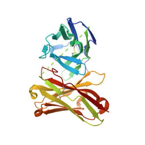
Three-dimensional structure of a high affinity anti-(4-hydroxy-3-nitrophenyl)acetyl antibody possessing a glycine residue at position 95 of the heavy chain.

Nishiguchi, A.Numoto, N.Ito, N.Azuma, T.Oda, M.

(2019) Mol Immunol 114: 545-552

  • DOI: https://doi.org/10.1016/j.molimm.2019.09.001
  • Primary Citation of Related Structures:  
    6K4Z

  • PubMed Abstract: 

    Antibodies possessing high affinity and specificity are desired as therapeutic reagents and biosensor materials. Such antibodies are often obtained from immunized animals through the process referred to as affinity maturation where antibody affinity increases with time after immunization. Somatic hypermutation (SHM) was shown to be involved in this process; however, structural basis of affinity maturation has not well been understood yet. We analyzed the crystal structure of a high affinity anti-(4-hydroxy-3-nitrophenyl)acetyl antibody, C6, possessing Gly at position 95 of heavy chain and 17 amino acid replacements by SHM. Here, we discuss how the amino acid residues at position 95, introduced at a junction of V H and D H gene segments during gene-recombination, as well as those replaced by SHM contribute to increasing the affinity by comparing the C6 structure with that of a germline low affinity antibody, N1G9, possessing Tyr at position 95.


  • Organizational Affiliation

    Graduate School of Life and Environmental Sciences, Kyoto Prefectural University, 1-5 Hangi-cho, Shimogamo, Sakyo-ku, Kyoto, 606-8522, Japan.


Macromolecules
Find similar proteins by:  (by identity cutoff)  |  3D Structure
Entity ID: 1
MoleculeChains Sequence LengthOrganismDetailsImage
scFv of C6
A, B
247Mus musculusMutation(s): 0 
Entity Groups  
Sequence Clusters30% Identity50% Identity70% Identity90% Identity95% Identity100% Identity
Sequence Annotations
Expand
  • Reference Sequence
Experimental Data & Validation

Experimental Data

  • Method: X-RAY DIFFRACTION
  • Resolution: 1.65 Å
  • R-Value Free: 0.196 
  • R-Value Work: 0.167 
  • R-Value Observed: 0.169 
  • Space Group: P 1
Unit Cell:
Length ( Å )Angle ( ˚ )
a = 37.393α = 88.235
b = 46.779β = 103.78
c = 74.581γ = 111.984
Software Package:
Software NamePurpose
PHENIXrefinement
Cootmodel building
PHASERphasing
XDSdata reduction
XDSdata scaling

Structure Validation

View Full Validation Report


Ligand Structure Quality Assessment 


Entry History 

Deposition Data

Revision History  (Full details and data files)

  • Version 1.0: 2019-09-18
    Type: Initial release
  • Version 1.1: 2019-10-02
    Changes: Data collection, Database references
  • Version 1.2: 2023-11-22
    Changes: Data collection, Database references, Refinement description