6JWC

Mouse antibody 2B5 Fab in complex with PEG


Experimental Data Snapshot

  • Method: X-RAY DIFFRACTION
  • Resolution: 2.30 Å
  • R-Value Free: 0.226 
  • R-Value Work: 0.187 
  • R-Value Observed: 0.189 

wwPDB Validation   3D Report Full Report


Ligand Structure Quality Assessment 


This is version 1.1 of the entry. See complete history


Literature

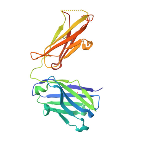
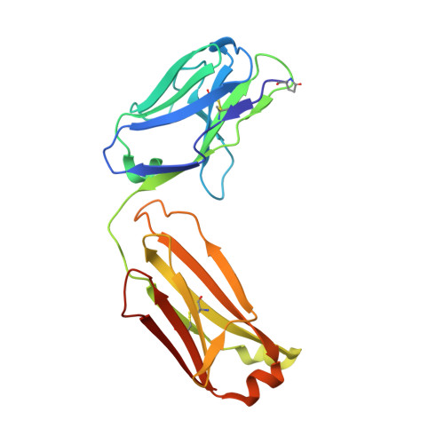
Structural basis of polyethylene glycol recognition by antibody.

Lee, C.C.Su, Y.C.Ko, T.P.Lin, L.L.Yang, C.Y.Chang, S.S.Roffler, S.R.Wang, A.H.

(2020) J Biomed Sci 27: 12-12

  • DOI: https://doi.org/10.1186/s12929-019-0589-7
  • Primary Citation of Related Structures:  
    6JP7, 6JU0, 6JWC

  • PubMed Abstract: 

    Polyethylene glycol (PEG) is widely used in industry and medicine. Anti-PEG antibodies have been developed for characterizing PEGylated drugs and other applications. However, the underlying mechanism for specific PEG binding has not been elucidated.


  • Organizational Affiliation

    Institute of Biological Chemistry, Academia Sinica, Taipei, Taiwan. chengung@gate.sinica.edu.tw.


Macromolecules
Find similar proteins by:  (by identity cutoff)  |  3D Structure
Entity ID: 1
MoleculeChains Sequence LengthOrganismDetailsImage
Immunoglobulin heavy chain Fab fragmentA [auth H]221Mus musculusMutation(s): 0 
Entity Groups  
Sequence Clusters30% Identity50% Identity70% Identity90% Identity95% Identity100% Identity
Sequence Annotations
Expand
  • Reference Sequence
Find similar proteins by:  (by identity cutoff)  |  3D Structure
Entity ID: 2
MoleculeChains Sequence LengthOrganismDetailsImage
Immunoglobulin light chainB [auth L]215Mus musculusMutation(s): 0 
Entity Groups  
Sequence Clusters30% Identity50% Identity70% Identity90% Identity95% Identity100% Identity
Sequence Annotations
Expand
  • Reference Sequence
Small Molecules
Ligands 4 Unique
IDChains Name / Formula / InChI Key2D Diagram3D Interactions
12P (Subject of Investigation/LOI)
Query on 12P

Download Ideal Coordinates CCD File 
G [auth H]DODECAETHYLENE GLYCOL
C24 H50 O13
WRZXKWFJEFFURH-UHFFFAOYSA-N
O4B
Query on O4B

Download Ideal Coordinates CCD File 
H,
I [auth H],
J [auth H],
M [auth L],
N [auth L]
1,4,7,10,13,16-HEXAOXACYCLOOCTADECANE
C12 H24 O6
XEZNGIUYQVAUSS-UHFFFAOYSA-N
PEG
Query on PEG

Download Ideal Coordinates CCD File 
L
DI(HYDROXYETHYL)ETHER
C4 H10 O3
MTHSVFCYNBDYFN-UHFFFAOYSA-N
SO4
Query on SO4

Download Ideal Coordinates CCD File 
C [auth H],
D [auth H],
E [auth H],
F [auth H],
K [auth L]
SULFATE ION
O4 S
QAOWNCQODCNURD-UHFFFAOYSA-L
Modified Residues  1 Unique
IDChains TypeFormula2D DiagramParent
PCA
Query on PCA
B [auth L]L-PEPTIDE LINKINGC5 H7 N O3GLN
Experimental Data & Validation

Experimental Data

  • Method: X-RAY DIFFRACTION
  • Resolution: 2.30 Å
  • R-Value Free: 0.226 
  • R-Value Work: 0.187 
  • R-Value Observed: 0.189 
  • Space Group: P 43 21 2
Unit Cell:
Length ( Å )Angle ( ˚ )
a = 98.895α = 90
b = 98.895β = 90
c = 96.711γ = 90
Software Package:
Software NamePurpose
PHENIXrefinement
HKL-2000data reduction
HKL-2000data scaling
MOLREPphasing

Structure Validation

View Full Validation Report


Ligand Structure Quality Assessment 


Entry History & Funding Information

Deposition Data


Funding OrganizationLocationGrant Number
Academia Sinica (Taiwan)Taiwan--

Revision History  (Full details and data files)

  • Version 1.0: 2020-02-05
    Type: Initial release
  • Version 1.1: 2023-11-22
    Changes: Data collection, Database references, Refinement description