6JNT

Catalase structure determined by eEFD (dataset 1)

  • Classification: OXIDOREDUCTASE
  • Organism(s): Bos taurus
  • Expression System: Bos taurus
  • Mutation(s): No 

  • Deposited: 2019-03-18 Released: 2019-04-24 
  • Deposition Author(s): Yonekura, K., Maki-Yonekura, S.
  • Funding Organization(s): Japan Society for the Promotion of Science, Japan Agency for Medical Research and Development (AMED), Japan Science and Technology

Experimental Data Snapshot

  • Method: ELECTRON CRYSTALLOGRAPHY
  • Resolution: 3.00 Å
  • R-Value Free: 0.283 
  • R-Value Work: 0.251 
  • R-Value Observed: 0.253 

wwPDB Validation   3D Report Full Report


This is version 1.2 of the entry. See complete history


Literature

A new cryo-EM system for electron 3D crystallography by eEFD.

Yonekura, K.Ishikawa, T.Maki-Yonekura, S.

(2019) J Struct Biol 206: 243-253

  • DOI: https://doi.org/10.1016/j.jsb.2019.03.009
  • Primary Citation of Related Structures:  
    6JNT, 6JNU

  • PubMed Abstract: 

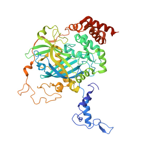
    A new cryo-EM system has been developed and investigated for use in protein electron 3D crystallography. The system provides parallel illumination of a coherent 300 kV electron beam to a sample, filters out energy-loss electrons through the sample with an in-column energy filter, and allows rotational data collection on a fast camera. It also possesses motorized cryo-sample loading and automated liquid-nitrogen filling for cooling of multiple samples. To facilitate its use, we developed GUI programs for efficient operation and accurate structure analysis. Here we report on the performance of the system and first results for thin 3D crystals of the protein complexes, catalase and membrane protein complex ExbBD. Data quality is remarkably improved with this approach, which we name eEFD (electron energy-filtered diffraction of 3D crystals), compared with those collected at 200 kV without energy filtration. Key advances include precise control of the microscope and recordings of lens fluctuations, which the programs process and respond to. We also discuss the merits of higher-energy electrons and filtration of energy-loss electrons in electron 3D crystallography.


  • Organizational Affiliation

    Biostructural Mechanism Laboratory, RIKEN, SPring-8 Center, 1-1-1 Kouto, Sayo, Hyogo 679-5148, Japan; Advanced Electron Microscope Development Unit, RIKEN-JEOL Collaboration Center, RIKEN Baton Zone Program, 1-1-1 Kouto, Sayo, Hyogo 679-5148, Japan. Electronic address: yone@spring8.or.jp.


Macromolecules
Find similar proteins by:  (by identity cutoff)  |  3D Structure
Entity ID: 1
MoleculeChains Sequence LengthOrganismDetailsImage
Catalase
A, B, C, D
527Bos taurusMutation(s): 0 
Gene Names: CAT
EC: 1.11.1.6
UniProt
Find proteins for P00432 (Bos taurus)
Explore P00432 
Go to UniProtKB:  P00432
Entity Groups  
Sequence Clusters30% Identity50% Identity70% Identity90% Identity95% Identity100% Identity
UniProt GroupP00432
Sequence Annotations
Expand
  • Reference Sequence
Experimental Data & Validation

Experimental Data

  • Method: ELECTRON CRYSTALLOGRAPHY
  • Resolution: 3.00 Å
  • R-Value Free: 0.283 
  • R-Value Work: 0.251 
  • R-Value Observed: 0.253 
  • Space Group: P 21 21 21
Unit Cell:
Length ( Å )Angle ( ˚ )
a = 69.109α = 90
b = 171.901β = 90
c = 195.013γ = 90
Software Package:
Software NamePurpose
PHENIXrefinement
EM Software:
TaskSoftware PackageVersion
MODEL REFINEMENTPHENIX1.14_3260

Structure Validation

View Full Validation Report


Entry History & Funding Information

Deposition Data


Funding OrganizationLocationGrant Number
Japan Society for the Promotion of ScienceJapan16H04757
Japan Society for the Promotion of ScienceJapan24657111
Japan Agency for Medical Research and Development (AMED)JapanCiCLE
Japan Science and TechnologyJapanSENTAN program

Revision History  (Full details and data files)

  • Version 1.0: 2019-04-24
    Type: Initial release
  • Version 1.1: 2019-05-22
    Changes: Data collection, Database references
  • Version 1.2: 2024-03-27
    Changes: Data collection, Database references, Refinement description