6JKI

Crystal structure and catalytic mechanism of the essential m1G37 tRNA methyltransferase TrmD from Pseudomonas aeruginosa


Experimental Data Snapshot

  • Method: X-RAY DIFFRACTION
  • Resolution: 2.59 Å
  • R-Value Free: 0.251 
  • R-Value Work: 0.207 
  • R-Value Observed: 0.209 

wwPDB Validation   3D Report Full Report


Ligand Structure Quality Assessment 


This is version 1.3 of the entry. See complete history


Literature

Crystal structure and catalytic mechanism of the essential m1G37 tRNA methyltransferase TrmD fromPseudomonas aeruginosa.

Jaroensuk, J.Wong, Y.H.Zhong, W.Liew, C.W.Maenpuen, S.Sahili, A.E.Atichartpongkul, S.Chionh, Y.H.Nah, Q.Thongdee, N.McBee, M.E.Prestwich, E.G.DeMott, M.S.Chaiyen, P.Mongkolsuk, S.Dedon, P.C.Lescar, J.Fuangthong, M.

(2019) RNA 25: 1481-1496

  • DOI: https://doi.org/10.1261/rna.066746.118
  • Primary Citation of Related Structures:  
    5WYQ, 5WYR, 6JKI

  • PubMed Abstract: 

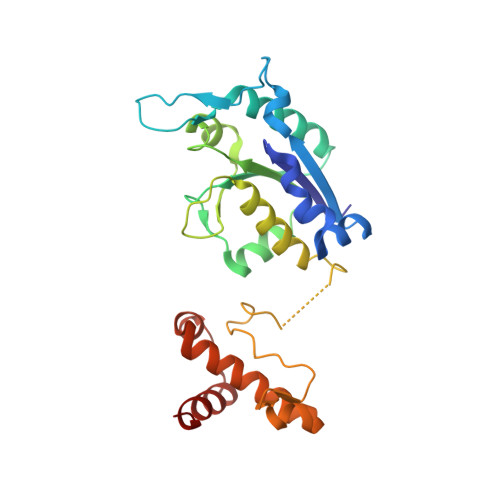
    The tRNA (m 1 G37) methyltransferase TrmD catalyzes m 1 G formation at position 37 in many tRNA isoacceptors and is essential in most bacteria, which positions it as a target for antibiotic development. In spite of its crucial role, little is known about TrmD in Pseudomonas aeruginosa ( Pa TrmD), an important human pathogen. Here we present detailed structural, substrate, and kinetic properties of Pa TrmD. The mass spectrometric analysis confirmed the G36G37-containing tRNAs Leu(GAG), Leu(CAG), Leu(UAG), Pro(GGG), Pro(UGG), Pro(CGG), and His(GUG) as Pa TrmD substrates. Analysis of steady-state kinetics with S -adenosyl-l-methionine (SAM) and tRNA Leu(GAG) showed that Pa TrmD catalyzes the two-substrate reaction by way of a ternary complex, while isothermal titration calorimetry revealed that SAM and tRNA Leu(GAG) bind to Pa TrmD independently, each with a dissociation constant of 14 ± 3 µM. Inhibition by the SAM analog sinefungin was competitive with respect to SAM ( K i = 0.41 ± 0.07 µM) and uncompetitive for tRNA ( K i = 6.4 ± 0.8 µM). A set of crystal structures of the homodimeric Pa TrmD protein bound to SAM and sinefungin provide the molecular basis for enzyme competitive inhibition and identify the location of the bound divalent ion. These results provide insights into Pa TrmD as a potential target for the development of antibiotics.


  • Organizational Affiliation

    Applied Biological Sciences Program, Chulabhorn Graduate Institute, Chulabhorn Royal Academy, Bangkok 10210, Thailand.


Macromolecules
Find similar proteins by:  (by identity cutoff)  |  3D Structure
Entity ID: 1
MoleculeChains Sequence LengthOrganismDetailsImage
tRNA (guanine-N(1)-)-methyltransferase
A, B
250Pseudomonas aeruginosa UCBPP-PA14Mutation(s): 0 
Gene Names: trmDPA14_15990
EC: 2.1.1.228
UniProt
Find proteins for Q9HXQ1 (Pseudomonas aeruginosa (strain ATCC 15692 / DSM 22644 / CIP 104116 / JCM 14847 / LMG 12228 / 1C / PRS 101 / PAO1))
Explore Q9HXQ1 
Go to UniProtKB:  Q9HXQ1
Entity Groups  
Sequence Clusters30% Identity50% Identity70% Identity90% Identity95% Identity100% Identity
UniProt GroupQ9HXQ1
Sequence Annotations
Expand
  • Reference Sequence
Small Molecules
Ligands 5 Unique
IDChains Name / Formula / InChI Key2D Diagram3D Interactions
SFG (Subject of Investigation/LOI)
Query on SFG

Download Ideal Coordinates CCD File 
C [auth A],
H [auth A]
SINEFUNGIN
C15 H23 N7 O5
LMXOHSDXUQEUSF-YECHIGJVSA-N
PEG
Query on PEG

Download Ideal Coordinates CCD File 
L [auth B],
P [auth B]
DI(HYDROXYETHYL)ETHER
C4 H10 O3
MTHSVFCYNBDYFN-UHFFFAOYSA-N
GOL
Query on GOL

Download Ideal Coordinates CCD File 
D [auth A]
E [auth A]
F [auth A]
G [auth A]
M [auth B]
D [auth A],
E [auth A],
F [auth A],
G [auth A],
M [auth B],
N [auth B],
O [auth B],
Q [auth B],
R [auth B],
S [auth B]
GLYCEROL
C3 H8 O3
PEDCQBHIVMGVHV-UHFFFAOYSA-N
MN (Subject of Investigation/LOI)
Query on MN

Download Ideal Coordinates CCD File 
I [auth A],
J [auth A],
T [auth B]
MANGANESE (II) ION
Mn
WAEMQWOKJMHJLA-UHFFFAOYSA-N
NA
Query on NA

Download Ideal Coordinates CCD File 
K [auth A],
U [auth B]
SODIUM ION
Na
FKNQFGJONOIPTF-UHFFFAOYSA-N
Experimental Data & Validation

Experimental Data

  • Method: X-RAY DIFFRACTION
  • Resolution: 2.59 Å
  • R-Value Free: 0.251 
  • R-Value Work: 0.207 
  • R-Value Observed: 0.209 
  • Space Group: P 32 2 1
Unit Cell:
Length ( Å )Angle ( ˚ )
a = 85.67α = 90
b = 85.67β = 90
c = 147.6γ = 120
Software Package:
Software NamePurpose
BUSTERrefinement
XDSdata reduction
XSCALEdata scaling
MOLREPphasing

Structure Validation

View Full Validation Report


Ligand Structure Quality Assessment 


Entry History 

Revision History  (Full details and data files)

  • Version 1.0: 2019-07-03
    Type: Initial release
  • Version 1.1: 2019-10-02
    Changes: Data collection, Database references
  • Version 1.2: 2019-10-30
    Changes: Data collection, Database references
  • Version 1.3: 2023-11-22
    Changes: Data collection, Database references, Derived calculations, Refinement description