6JF1

Crystal structure of the substrate binding protein of a methionine transporter from Streptococcus pneumoniae


Experimental Data Snapshot

  • Method: X-RAY DIFFRACTION
  • Resolution: 1.95 Å
  • R-Value Free: 0.231 
  • R-Value Work: 0.183 
  • R-Value Observed: 0.186 

wwPDB Validation   3D Report Full Report


This is version 1.2 of the entry. See complete history


Literature

Structural insights into the substrate specificity of SP_0149, the substrate-binding protein of a methionine ABC transporter from Streptococcus pneumoniae.

Jha, B.Vyas, R.Bhushan, J.Sehgal, D.Biswal, B.K.

(2019) Acta Crystallogr F Struct Biol Commun 75: 520-528

  • DOI: https://doi.org/10.1107/S2053230X19009038
  • Primary Citation of Related Structures:  
    6JF1

  • PubMed Abstract: 

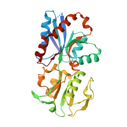
    Successful pathogenesis is a cumulative effect of the virulence factors of a pathogen and its capability to efficiently utilize the available nutrients from the host. Streptococcus pneumoniae, a Gram-positive opportunistic pathogen, may either reside asymptomatically as a nasopharyngeal commensal inside the human host or cause lethal diseases, including pneumonia, meningitis and sepsis. S. pneumoniae is known to acquire methionine (Met) from its host through a Met importer. Here, the crystal structure of the substrate-binding protein (SBP; SP_0149) of an ABC importer with Met bound is reported at a resolution of 1.95 Å. The three-dimensional structure of SBP shows that it is composed of two distinct domains, each consisting of a mixed β-sheet flanked by helices. The substrate, Met, is bound in the central part of the interface between the two domains. The overall structure of SP_0149 resembles those of SBPs from other reported bacterial Met and Gly-Met dipeptide transporters. However, a detailed analysis of these structures shows notable variations in the amino-acid composition of the substrate-binding pockets of the SP_0149-Met and GmpC-Gly-Met structures. In particular, SP_0149 harbors Thr212 and Tyr114, whereas the corresponding residues in GmpC are Gly and Val. This difference is likely to be the underlying basis for their differential substrate specificity. In summary, the structure of the SP_0149-Met complex provides insights into the transport function of SP_0149 and its interactions with methionine. It opens up avenues for the rational design of inhibitors of SP_0149 through a structure-mediated approach.


  • Organizational Affiliation

    Structural and Functional Biology Laboratory, National Institute of Immunology, Aruna Asaf Ali Marg, New Delhi 110 067, India.


Macromolecules
Find similar proteins by:  (by identity cutoff)  |  3D Structure
Entity ID: 1
MoleculeChains Sequence LengthOrganismDetailsImage
Lipoprotein276Streptococcus pneumoniae TIGR4Mutation(s): 0 
Gene Names: SP_0149
UniProt
Find proteins for A0A0H2UN92 (Streptococcus pneumoniae serotype 4 (strain ATCC BAA-334 / TIGR4))
Explore A0A0H2UN92 
Go to UniProtKB:  A0A0H2UN92
Entity Groups  
Sequence Clusters30% Identity50% Identity70% Identity90% Identity95% Identity100% Identity
UniProt GroupA0A0H2UN92
Sequence Annotations
Expand
  • Reference Sequence
Small Molecules
Ligands 5 Unique
IDChains Name / Formula / InChI Key2D Diagram3D Interactions
MET
Query on MET

Download Ideal Coordinates CCD File 
B [auth A]METHIONINE
C5 H11 N O2 S
FFEARJCKVFRZRR-BYPYZUCNSA-N
GOL
Query on GOL

Download Ideal Coordinates CCD File 
W [auth A],
X [auth A]
GLYCEROL
C3 H8 O3
PEDCQBHIVMGVHV-UHFFFAOYSA-N
ZN
Query on ZN

Download Ideal Coordinates CCD File 
C [auth A]
D [auth A]
E [auth A]
F [auth A]
G [auth A]
C [auth A],
D [auth A],
E [auth A],
F [auth A],
G [auth A],
H [auth A],
I [auth A],
J [auth A],
K [auth A],
L [auth A],
M [auth A]
ZINC ION
Zn
PTFCDOFLOPIGGS-UHFFFAOYSA-N
ACT
Query on ACT

Download Ideal Coordinates CCD File 
O [auth A]
P [auth A]
Q [auth A]
R [auth A]
S [auth A]
O [auth A],
P [auth A],
Q [auth A],
R [auth A],
S [auth A],
T [auth A],
U [auth A],
V [auth A]
ACETATE ION
C2 H3 O2
QTBSBXVTEAMEQO-UHFFFAOYSA-M
CL
Query on CL

Download Ideal Coordinates CCD File 
N [auth A]CHLORIDE ION
Cl
VEXZGXHMUGYJMC-UHFFFAOYSA-M
Experimental Data & Validation

Experimental Data

  • Method: X-RAY DIFFRACTION
  • Resolution: 1.95 Å
  • R-Value Free: 0.231 
  • R-Value Work: 0.183 
  • R-Value Observed: 0.186 
  • Space Group: P 21 21 21
Unit Cell:
Length ( Å )Angle ( ˚ )
a = 54.692α = 90
b = 75.851β = 90
c = 75.634γ = 90
Software Package:
Software NamePurpose
REFMACrefinement
HKL-2000data reduction
HKL-2000data scaling
PHASERphasing

Structure Validation

View Full Validation Report


Entry History & Funding Information

Deposition Data


Funding OrganizationLocationGrant Number
Department of Biotechnology (India)India--

Revision History  (Full details and data files)

  • Version 1.0: 2019-07-10
    Type: Initial release
  • Version 1.1: 2021-01-27
    Changes: Database references, Derived calculations
  • Version 1.2: 2023-11-22
    Changes: Data collection, Database references, Refinement description