6HUF

Coping with strong translational non-crystallographic symmetry and extreme anisotropy in molecular replacement with Phaser: human Rab27a


Experimental Data Snapshot

  • Method: X-RAY DIFFRACTION
  • Resolution: 2.82 Å
  • R-Value Free: 0.342 
  • R-Value Work: 0.312 
  • R-Value Observed: 0.313 

wwPDB Validation   3D Report Full Report


Ligand Structure Quality Assessment 


This is version 1.3 of the entry. See complete history


Literature

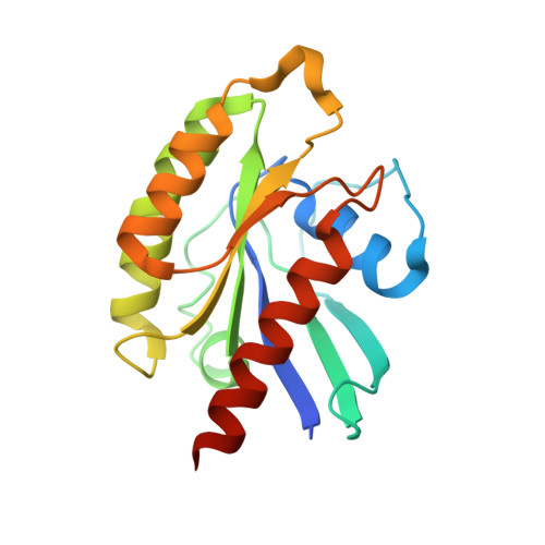
Coping with strong translational noncrystallographic symmetry and extreme anisotropy in molecular replacement with Phaser: human Rab27a.

Jamshidiha, M.Perez-Dorado, I.Murray, J.W.Tate, E.W.Cota, E.Read, R.J.

(2019) Acta Crystallogr D Struct Biol 75: 342-353

  • DOI: https://doi.org/10.1107/S2059798318017825
  • Primary Citation of Related Structures:  
    6HUF

  • PubMed Abstract: 

    Data pathologies caused by effects such as diffraction anisotropy and translational noncrystallographic symmetry (tNCS) can dramatically complicate the solution of the crystal structures of macromolecules. Such problems were encountered in determining the structure of a mutant form of Rab27a, a member of the Rab GTPases. Mutant Rab27a constructs that crystallize in the free form were designed for use in the discovery of drugs to reduce primary tumour invasiveness and metastasis. One construct, hRab27a Mut , crystallized within 24 h and diffracted to 2.82 Å resolution, with a unit cell possessing room for a large number of protein copies. Initial efforts to solve the structure using molecular replacement by Phaser were not successful. Analysis of the data set revealed that the crystals suffered from both extreme anisotropy and strong tNCS. As a result, large numbers of reflections had estimated standard deviations that were much larger than their measured intensities and their expected intensities, revealing problems with the use of such data at the time in Phaser. By eliminating extremely weak reflections with the largest combined effects of anisotropy and tNCS, these problems could be avoided, allowing a molecular-replacement solution to be found. The lessons that were learned in solving this structure have guided improvements in the numerical analysis used in Phaser, particularly in identifying diffraction measurements that convey very little information content. The calculation of information content could also be applied as an alternative to ellipsoidal truncation. The post-mortem analysis also revealed an oversight in accounting for measurement errors in the fast rotation function. While the crystal of mutant Rab27a is not amenable to drug screening, the structure can guide new modifications to obtain more suitable crystal forms.


  • Organizational Affiliation

    Faculty of Natural Sciences, Department of Life Sciences, Imperial College London, Exhibition Road, South Kensington, London SW7 2AZ, England.


Macromolecules
Find similar proteins by:  (by identity cutoff)  |  3D Structure
Entity ID: 1
MoleculeChains Sequence LengthOrganismDetailsImage
Ras-related protein Rab-27A182Homo sapiensMutation(s): 0 
Gene Names: RAB27ARAB27
UniProt & NIH Common Fund Data Resources
Find proteins for P51159 (Homo sapiens)
Explore P51159 
Go to UniProtKB:  P51159
PHAROS:  P51159
GTEx:  ENSG00000069974 
Entity Groups  
Sequence Clusters30% Identity50% Identity70% Identity90% Identity95% Identity100% Identity
UniProt GroupP51159
Sequence Annotations
Expand
  • Reference Sequence
Small Molecules
Ligands 2 Unique
IDChains Name / Formula / InChI Key2D Diagram3D Interactions
GNP
Query on GNP

Download Ideal Coordinates CCD File 
BA [auth C]
DA [auth K]
FA [auth M]
HA [auth D]
JA [auth P]
BA [auth C],
DA [auth K],
FA [auth M],
HA [auth D],
JA [auth P],
LA [auth F],
NA [auth J],
PA [auth G],
R [auth A],
RA [auth I],
T [auth O],
TA [auth H],
V [auth N],
VA [auth L],
X [auth B],
Z [auth E]
PHOSPHOAMINOPHOSPHONIC ACID-GUANYLATE ESTER
C10 H17 N6 O13 P3
UQABYHGXWYXDTK-UUOKFMHZSA-N
MG
Query on MG

Download Ideal Coordinates CCD File 
AA [auth C]
CA [auth K]
EA [auth M]
GA [auth D]
IA [auth P]
AA [auth C],
CA [auth K],
EA [auth M],
GA [auth D],
IA [auth P],
KA [auth F],
MA [auth J],
OA [auth G],
Q [auth A],
QA [auth I],
S [auth O],
SA [auth H],
U [auth N],
UA [auth L],
W [auth B],
Y [auth E]
MAGNESIUM ION
Mg
JLVVSXFLKOJNIY-UHFFFAOYSA-N
Experimental Data & Validation

Experimental Data

  • Method: X-RAY DIFFRACTION
  • Resolution: 2.82 Å
  • R-Value Free: 0.342 
  • R-Value Work: 0.312 
  • R-Value Observed: 0.313 
  • Space Group: C 1 2 1
Unit Cell:
Length ( Å )Angle ( ˚ )
a = 130.42α = 90
b = 132.41β = 103.57
c = 230.59γ = 90
Software Package:
Software NamePurpose
REFMACrefinement
DIALSdata reduction
DIALSdata scaling
PHASERphasing

Structure Validation

View Full Validation Report


Ligand Structure Quality Assessment 


Entry History & Funding Information

Deposition Data


Funding OrganizationLocationGrant Number
Wellcome TrustUnited Kingdom082961/Z/07/Z
Cancer Research UKUnited KingdomC29637/A20781

Revision History  (Full details and data files)

  • Version 1.0: 2019-03-27
    Type: Initial release
  • Version 1.1: 2019-04-17
    Changes: Data collection, Database references
  • Version 1.2: 2019-07-10
    Changes: Data collection
  • Version 1.3: 2024-01-24
    Changes: Data collection, Database references, Derived calculations, Refinement description