6HIG

hPD-1/NBO1a Fab complex


Experimental Data Snapshot

  • Method: X-RAY DIFFRACTION
  • Resolution: 2.20 Å
  • R-Value Free: 0.270 
  • R-Value Work: 0.215 
  • R-Value Observed: 0.217 

wwPDB Validation   3D Report Full Report


This is version 1.2 of the entry. See complete history


Literature

Tumor suppression of novel anti-PD-1 antibodies mediated through CD28 costimulatory pathway.

Fenwick, C.Loredo-Varela, J.L.Joo, V.Pellaton, C.Farina, A.Rajah, N.Esteves-Leuenberger, L.Decaillon, T.Suffiotti, M.Noto, A.Ohmiti, K.Gottardo, R.Weissenhorn, W.Pantaleo, G.

(2019) J Exp Med 216: 1525-1541

  • DOI: https://doi.org/10.1084/jem.20182359
  • Primary Citation of Related Structures:  
    6HIG

  • PubMed Abstract: 

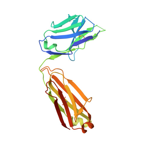
    Classical antagonistic antibodies (Abs) targeting PD-1, such as pembrolizumab and nivolumab, act through blockade of the PD-1-PDL-1 interaction. Here, we have identified novel antagonistic anti-PD-1 Abs not blocking the PD-1-PDL-1 interaction. The nonblocking Abs recognize epitopes on PD-1 located on the opposing face of the PDL-1 interaction and overlap with a newly identified evolutionarily conserved patch. These nonblocking Abs act predominantly through the CD28 coreceptor. Importantly, a combination of blocking and nonblocking Abs synergize in the functional recovery of antigen-specific exhausted CD8 T cells. Interestingly, nonblocking anti-PD-1 Abs have equivalent antitumor activity compared with blocker Abs in two mouse tumor models, and combination therapy using both classes of Abs enhanced tumor suppression in the mouse immunogenic tumor model. The identification of the novel nonblocker anti-PD-1 Abs and their synergy with classical blocker Abs may be instrumental in potentiating immunotherapy strategies and antitumor activity.


  • Organizational Affiliation

    Service of Immunology and Allergy, Department of Medicine, Lausanne University Hospital, University of Lausanne, Lausanne, Switzerland.


Macromolecules
Find similar proteins by:  (by identity cutoff)  |  3D Structure
Entity ID: 1
MoleculeChains Sequence LengthOrganismDetailsImage
Heavy ChainA [auth H]233Mus musculusMutation(s): 0 
Entity Groups  
Sequence Clusters30% Identity50% Identity70% Identity90% Identity95% Identity100% Identity
Sequence Annotations
Expand
  • Reference Sequence
Find similar proteins by:  (by identity cutoff)  |  3D Structure
Entity ID: 2
MoleculeChains Sequence LengthOrganismDetailsImage
Light ChainB [auth L]214Mus musculusMutation(s): 0 
Entity Groups  
Sequence Clusters30% Identity50% Identity70% Identity90% Identity95% Identity100% Identity
Sequence Annotations
Expand
  • Reference Sequence
Find similar proteins by:  (by identity cutoff)  |  3D Structure
Entity ID: 3
MoleculeChains Sequence LengthOrganismDetailsImage
Programmed cell death protein 1C [auth B]118Homo sapiensMutation(s): 1 
Gene Names: PDCD1PD1
UniProt & NIH Common Fund Data Resources
Find proteins for Q15116 (Homo sapiens)
Explore Q15116 
Go to UniProtKB:  Q15116
PHAROS:  Q15116
GTEx:  ENSG00000188389 
Entity Groups  
Sequence Clusters30% Identity50% Identity70% Identity90% Identity95% Identity100% Identity
UniProt GroupQ15116
Sequence Annotations
Expand
  • Reference Sequence
Experimental Data & Validation

Experimental Data

  • Method: X-RAY DIFFRACTION
  • Resolution: 2.20 Å
  • R-Value Free: 0.270 
  • R-Value Work: 0.215 
  • R-Value Observed: 0.217 
  • Space Group: P 1 21 1
Unit Cell:
Length ( Å )Angle ( ˚ )
a = 64.514α = 90
b = 63.049β = 106.49
c = 68.297γ = 90
Software Package:
Software NamePurpose
PHENIXrefinement
XDSdata reduction
Aimlessdata scaling
PHASERphasing

Structure Validation

View Full Validation Report


Entry History 

Deposition Data

Revision History  (Full details and data files)

  • Version 1.0: 2019-06-05
    Type: Initial release
  • Version 1.1: 2019-07-10
    Changes: Data collection, Database references
  • Version 1.2: 2024-01-17
    Changes: Data collection, Database references, Refinement description