An Aspartate-Specific Solute-Binding Protein Regulates Protein Kinase G Activity To Control Glutamate Metabolism in Mycobacteria.
Bhattacharyya, N., Nkumama, I.N., Newland-Smith, Z., Lin, L.Y., Yin, W., Cullen, R.E., Griffiths, J.S., Jarvis, A.R., Price, M.J., Chong, P.Y., Wallis, R., O'Hare, H.M.(2018) mBio 9
- PubMed: 30065086
- DOI: https://doi.org/10.1128/mBio.00931-18
- Primary Citation of Related Structures:
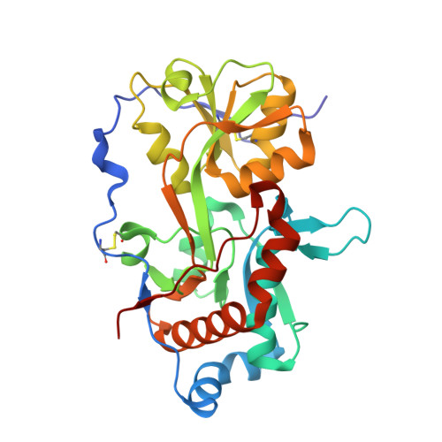
6H1U, 6H20, 6H2T - PubMed Abstract:
Signaling by serine/threonine phosphorylation controls diverse processes in bacteria, and identification of the stimuli that activate protein kinases is an outstanding question in the field. Recently, we showed that nutrients stimulate phosphorylation of the protein kinase G substrate GarA in Mycobacterium smegmatis and Mycobacterium tuberculosis and that the action of GarA in regulating central metabolism depends upon whether it is phosphorylated. Here we present an investigation into the mechanism by which nutrients activate PknG. Two unknown genes were identified as co-conserved and co-expressed with PknG: their products were a putative lipoprotein, GlnH, and putative transmembrane protein, GlnX. Using a genetic approach, we showed that the membrane protein GlnX is functionally linked to PknG. Furthermore, we determined that the ligand specificity of GlnH matches the amino acids that stimulate GarA phosphorylation. We determined the structure of GlnH in complex with different amino acid ligands (aspartate, glutamate, and asparagine), revealing the structural basis of ligand specificity. We propose that the amino acid concentration in the periplasm is sensed by GlnH and that protein-protein interaction allows transmission of this information across the membrane via GlnX to activate PknG. This sensory system would allow regulation of nutrient utilization in response to changes in nutrient availability. The sensor, signaling, and effector proteins are conserved throughout the Actinobacteria , including the important human pathogen Mycobacterium tuberculosis , industrial amino acid producer Corynebacterium glutamicum , and antibiotic-producing Streptomyces species. IMPORTANCE Tuberculosis (TB) kills 5,000 people every day, and the prevalence of multidrug-resistant TB is increasing in every country. The processes by which the pathogen Mycobacterium tuberculosis senses and responds to changes in its environment are attractive targets for drug development. Bacterial metabolism differs dramatically between growing and dormant cells, and these changes are known to be important in pathogenesis of TB. Here, we used genetic and biochemical approaches to identify proteins that allow M. tuberculosis to detect amino acids in its surroundings so that it can regulate its metabolism. We have also shown how individual amino acids are recognized. The findings have broader significance for other actinobacterial pathogens, such as nontuberculous mycobacteria, as well as Actinobacteria used to produce billions of dollars of amino acids and antibiotics every year.
Organizational Affiliation:
Leicester Tuberculosis Research Group, Department of Infection, Immunity and Inflammation, University of Leicester, Leicester, United Kingdom.