6GH4

HLA-E*01:03 in complex with the Mtb44 peptide variant: Mtb44*P2-Gln.


Experimental Data Snapshot

  • Method: X-RAY DIFFRACTION
  • Resolution: 2.16 Å
  • R-Value Free: 0.242 
  • R-Value Work: 0.202 
  • R-Value Observed: 0.204 

wwPDB Validation   3D Report Full Report


This is version 1.2 of the entry. See complete history


Literature

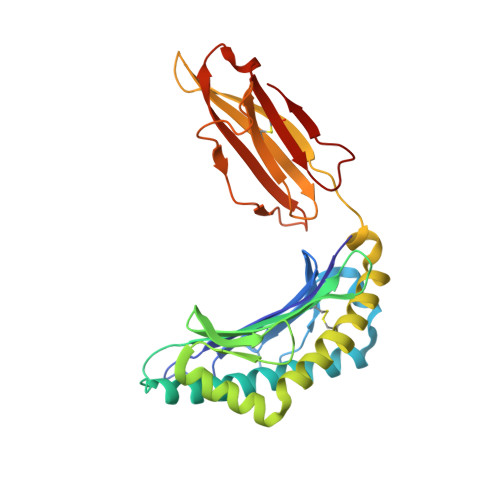
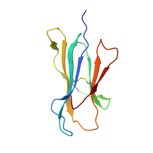
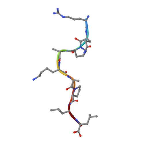
Pathogen-derived HLA-E bound epitopes reveal broad primary anchor pocket tolerability and conformationally malleable peptide binding.

Walters, L.C.Harlos, K.Brackenridge, S.Rozbesky, D.Barrett, J.R.Jain, V.Walter, T.S.O'Callaghan, C.A.Borrow, P.Toebes, M.Hansen, S.G.Sacha, J.Abdulhaqq, S.Greene, J.M.Fruh, K.Marshall, E.Picker, L.J.Jones, E.Y.McMichael, A.J.Gillespie, G.M.

(2018) Nat Commun 9: 3137-3137

  • DOI: https://doi.org/10.1038/s41467-018-05459-z
  • Primary Citation of Related Structures:  
    6GGM, 6GH1, 6GH4, 6GHN, 6GL1

  • PubMed Abstract: 

    Through major histocompatibility complex class Ia leader sequence-derived (VL9) peptide binding and CD94/NKG2 receptor engagement, human leucocyte antigen E (HLA-E) reports cellular health to NK cells. Previous studies demonstrated a strong bias for VL9 binding by HLA-E, a preference subsequently supported by structural analyses. However, Mycobacteria tuberculosis (Mtb) infection and Rhesus cytomegalovirus-vectored SIV vaccinations revealed contexts where HLA-E and the rhesus homologue, Mamu-E, presented diverse pathogen-derived peptides to CD8 + T cells, respectively. Here we present crystal structures of HLA-E in complex with HIV and Mtb-derived peptides. We show that despite the presence of preferred primary anchor residues, HLA-E-bound peptides can adopt alternative conformations within the peptide binding groove. Furthermore, combined structural and mutagenesis analyses illustrate a greater tolerance for hydrophobic and polar residues in the primary pockets than previously appreciated. Finally, biochemical studies reveal HLA-E peptide binding and exchange characteristics with potential relevance to its alternative antigen presenting function in vivo.


  • Organizational Affiliation

    Nuffield Department of Medicine Research Building, Roosevelt Drive, Nuffield Department of Medicine, University of Oxford, Oxford, OX3 7FZ, UK.


Macromolecules
Find similar proteins by:  (by identity cutoff)  |  3D Structure
Entity ID: 1
MoleculeChains Sequence LengthOrganismDetailsImage
MHC class I antigen
A, C, E, G
274Homo sapiensMutation(s): 0 
Gene Names: HLA-E
UniProt & NIH Common Fund Data Resources
Find proteins for P13747 (Homo sapiens)
Explore P13747 
Go to UniProtKB:  P13747
PHAROS:  P13747
GTEx:  ENSG00000204592 
Entity Groups  
Sequence Clusters30% Identity50% Identity70% Identity90% Identity95% Identity100% Identity
UniProt GroupP13747
Sequence Annotations
Expand
  • Reference Sequence
Find similar proteins by:  (by identity cutoff)  |  3D Structure
Entity ID: 2
MoleculeChains Sequence LengthOrganismDetailsImage
Beta-2-microglobulin
B, D, F, H
100Homo sapiensMutation(s): 0 
Gene Names: B2MCDABP0092HDCMA22P
UniProt & NIH Common Fund Data Resources
Find proteins for P61769 (Homo sapiens)
Explore P61769 
Go to UniProtKB:  P61769
PHAROS:  P61769
GTEx:  ENSG00000166710 
Entity Groups  
Sequence Clusters30% Identity50% Identity70% Identity90% Identity95% Identity100% Identity
UniProt GroupP61769
Sequence Annotations
Expand
  • Reference Sequence

Find similar proteins by:  Sequence   |   3D Structure  

Entity ID: 3
MoleculeChains Sequence LengthOrganismDetailsImage
ARG-GLN-PRO-ALA-LYS-ALA-PRO-LEU-LEUI [auth P],
J [auth Q],
K [auth R],
L [auth Y]
9MycobacteriaceaeMutation(s): 0 
UniProt
Find proteins for P9WGR1 (Mycobacterium tuberculosis (strain ATCC 25618 / H37Rv))
Explore P9WGR1 
Go to UniProtKB:  P9WGR1
Entity Groups  
UniProt GroupP9WGR1
Sequence Annotations
Expand
  • Reference Sequence
Experimental Data & Validation

Experimental Data

  • Method: X-RAY DIFFRACTION
  • Resolution: 2.16 Å
  • R-Value Free: 0.242 
  • R-Value Work: 0.202 
  • R-Value Observed: 0.204 
  • Space Group: P 1
Unit Cell:
Length ( Å )Angle ( ˚ )
a = 48.317α = 99.39
b = 76.412β = 99.83
c = 111.309γ = 93.5
Software Package:
Software NamePurpose
PHENIXrefinement
xia2data reduction
xia2data scaling
MOLREPphasing
Cootmodel building

Structure Validation

View Full Validation Report


Entry History & Funding Information

Deposition Data


Funding OrganizationLocationGrant Number
Bill & Melinda Gates FoundationUnited StatesOPP1133649

Revision History  (Full details and data files)

  • Version 1.0: 2018-08-08
    Type: Initial release
  • Version 1.1: 2018-08-22
    Changes: Data collection, Database references
  • Version 1.2: 2024-01-17
    Changes: Data collection, Database references, Derived calculations, Refinement description