6GB7

Structure of H-2Db with scoop loop from tapasin


Experimental Data Snapshot

  • Method: X-RAY DIFFRACTION
  • Resolution: 2.15 Å
  • R-Value Free: 0.266 
  • R-Value Work: 0.239 
  • R-Value Observed: 0.240 

wwPDB Validation   3D Report Full Report


This is version 1.3 of the entry. See complete history


Literature

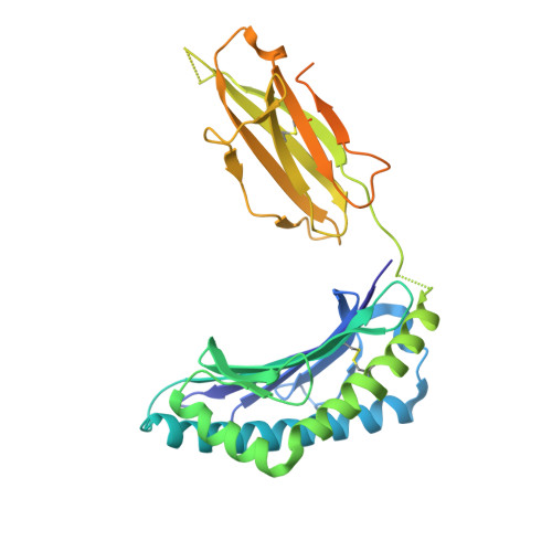
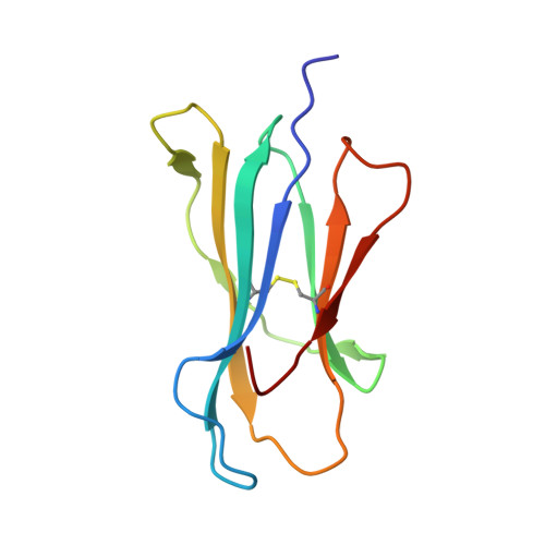
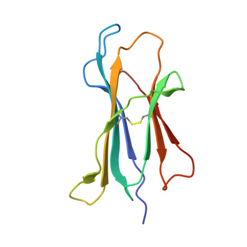
Successive crystal structure snapshots suggest the basis for MHC class I peptide loading and editing by tapasin.

Hafstrand, I.Sayitoglu, E.C.Apavaloaei, A.Josey, B.J.Sun, R.Han, X.Pellegrino, S.Ozkazanc, D.Potens, R.Janssen, L.Nilvebrant, J.Nygren, P.A.Sandalova, T.Springer, S.Georgoudaki, A.M.Duru, A.D.Achour, A.

(2019) Proc Natl Acad Sci U S A 116: 5055-5060

  • DOI: https://doi.org/10.1073/pnas.1807656116
  • Primary Citation of Related Structures:  
    6GB5, 6GB6, 6GB7

  • PubMed Abstract: 

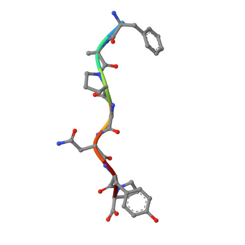
    MHC-I epitope presentation to CD8 + T cells is directly dependent on peptide loading and selection during antigen processing. However, the exact molecular bases underlying peptide selection and binding by MHC-I remain largely unknown. Within the peptide-loading complex, the peptide editor tapasin is key to the selection of MHC-I-bound peptides. Here, we have determined an ensemble of crystal structures of MHC-I in complex with the peptide exchange-associated dipeptide GL, as well as the tapasin-associated scoop loop, alone or in combination with candidate epitopes. These results combined with mutation analyses allow us to propose a molecular model underlying MHC-I peptide selection by tapasin. The N termini of bound peptides most probably bind first in the N-terminal and middle region of the MHC-I peptide binding cleft, upon which the peptide C termini are tested for their capacity to dislodge the tapasin scoop loop from the F pocket of the MHC-I cleft. Our results also indicate important differences in peptide selection between different MHC-I alleles.


  • Organizational Affiliation

    Science for Life Laboratory, Department of Medicine Solna, Karolinska Institute, and Division of Infectious Diseases, Karolinska University Hospital, Solna, SE-17176 Stockholm, Sweden.


Macromolecules
Find similar proteins by:  (by identity cutoff)  |  3D Structure
Entity ID: 1
MoleculeChains Sequence LengthOrganismDetailsImage
H-2 class I histocompatibility antigen, D-B alpha chain
A, C, E, G
338Mus musculusMutation(s): 0 
Gene Names: H2-D1
UniProt
Find proteins for P01899 (Mus musculus)
Explore P01899 
Go to UniProtKB:  P01899
Entity Groups  
Sequence Clusters30% Identity50% Identity70% Identity90% Identity95% Identity100% Identity
UniProt GroupP01899
Sequence Annotations
Expand
  • Reference Sequence
Find similar proteins by:  (by identity cutoff)  |  3D Structure
Entity ID: 2
MoleculeChains Sequence LengthOrganismDetailsImage
Beta-2-microglobulin
B, D, H
100Mus musculusMutation(s): 1 
Gene Names: B2m
UniProt & NIH Common Fund Data Resources
Find proteins for P01887 (Mus musculus)
Explore P01887 
Go to UniProtKB:  P01887
IMPC:  MGI:88127
Entity Groups  
Sequence Clusters30% Identity50% Identity70% Identity90% Identity95% Identity100% Identity
UniProt GroupP01887
Sequence Annotations
Expand
  • Reference Sequence
Find similar proteins by:  (by identity cutoff)  |  3D Structure
Entity ID: 3
MoleculeChains Sequence LengthOrganismDetailsImage
Beta-2-microglobulin99Mus musculusMutation(s): 1 
Gene Names: B2m
UniProt & NIH Common Fund Data Resources
Find proteins for P01887 (Mus musculus)
Explore P01887 
Go to UniProtKB:  P01887
IMPC:  MGI:88127
Entity Groups  
Sequence Clusters30% Identity50% Identity70% Identity90% Identity95% Identity100% Identity
UniProt GroupP01887
Sequence Annotations
Expand
  • Reference Sequence

Find similar proteins by:  Sequence   |   3D Structure  

Entity ID: 4
MoleculeChains Sequence LengthOrganismDetailsImage
PHE-ALA-PRO-GLY-ASN-TYR-PRO
I, J, K, L
7Respirovirus murisMutation(s): 0 
UniProt
Find proteins for O57286 (Sendai virus (strain Ohita))
Explore O57286 
Go to UniProtKB:  O57286
Entity Groups  
UniProt GroupO57286
Sequence Annotations
Expand
  • Reference Sequence

Find similar proteins by:  Sequence   |   3D Structure  

Entity ID: 5
MoleculeChains Sequence LengthOrganismDetailsImage
GLY-GLY-LEU-SERM [auth P],
N [auth R]
10Mus musculusMutation(s): 0 
UniProt
Find proteins for Q9R233 (Mus musculus)
Explore Q9R233 
Go to UniProtKB:  Q9R233
Entity Groups  
Sequence Clusters30% Identity50% Identity70% Identity90% Identity95% Identity100% Identity
UniProt GroupQ9R233
Sequence Annotations
Expand
  • Reference Sequence
Small Molecules
Ligands 2 Unique
IDChains Name / Formula / InChI Key2D Diagram3D Interactions
SO4
Query on SO4

Download Ideal Coordinates CCD File 
AA [auth G],
O [auth A],
W [auth C],
Z [auth F]
SULFATE ION
O4 S
QAOWNCQODCNURD-UHFFFAOYSA-L
GOL
Query on GOL

Download Ideal Coordinates CCD File 
BA [auth G]
CA [auth G]
P [auth A]
Q [auth A]
R [auth A]
BA [auth G],
CA [auth G],
P [auth A],
Q [auth A],
R [auth A],
S [auth A],
T [auth B],
U [auth B],
V [auth B],
X [auth C],
Y [auth D]
GLYCEROL
C3 H8 O3
PEDCQBHIVMGVHV-UHFFFAOYSA-N
Experimental Data & Validation

Experimental Data

  • Method: X-RAY DIFFRACTION
  • Resolution: 2.15 Å
  • R-Value Free: 0.266 
  • R-Value Work: 0.239 
  • R-Value Observed: 0.240 
  • Space Group: P 1 21 1
Unit Cell:
Length ( Å )Angle ( ˚ )
a = 92.7α = 90
b = 123.86β = 103.66
c = 100.11γ = 90
Software Package:
Software NamePurpose
PHENIXrefinement
XDSdata reduction
Aimlessdata scaling
PHASERphasing

Structure Validation

View Full Validation Report


Entry History 

Deposition Data

Revision History  (Full details and data files)

  • Version 1.0: 2019-03-06
    Type: Initial release
  • Version 1.1: 2019-03-13
    Changes: Data collection, Database references
  • Version 1.2: 2019-03-20
    Changes: Data collection, Database references
  • Version 1.3: 2024-01-17
    Changes: Data collection, Database references, Refinement description