6FZQ

Crystal structure of scFv-SM3 in complex with compound 3


Experimental Data Snapshot

  • Method: X-RAY DIFFRACTION
  • Resolution: 1.70 Å
  • R-Value Free: 0.225 
  • R-Value Work: 0.175 
  • R-Value Observed: 0.177 

wwPDB Validation   3D Report Full Report


Ligand Structure Quality Assessment 


This is version 2.1 of the entry. See complete history


Literature

Water Sculpts the Distinctive Shapes and Dynamics of the Tumor-Associated Carbohydrate Tn Antigens: Implications for Their Molecular Recognition.

Bermejo, I.A.Usabiaga, I.Companon, I.Castro-Lopez, J.Insausti, A.Fernandez, J.A.Avenoza, A.Busto, J.H.Jimenez-Barbero, J.Asensio, J.L.Peregrina, J.M.Jimenez-Oses, G.Hurtado-Guerrero, R.Cocinero, E.J.Corzana, F.

(2018) J Am Chem Soc 140: 9952-9960

  • DOI: https://doi.org/10.1021/jacs.8b04801
  • Primary Citation of Related Structures:  
    6FZQ, 6FZR

  • PubMed Abstract: 

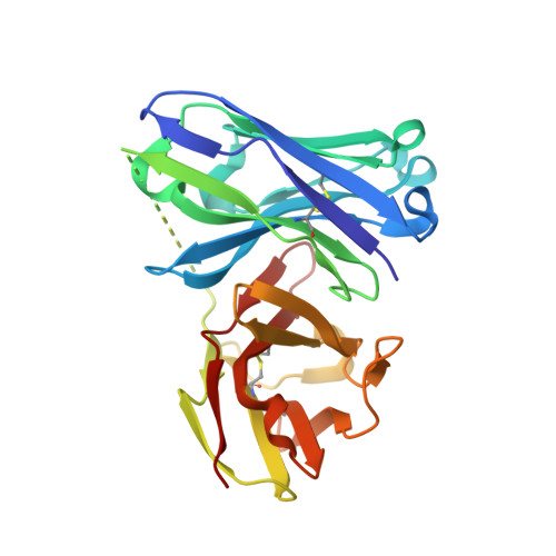
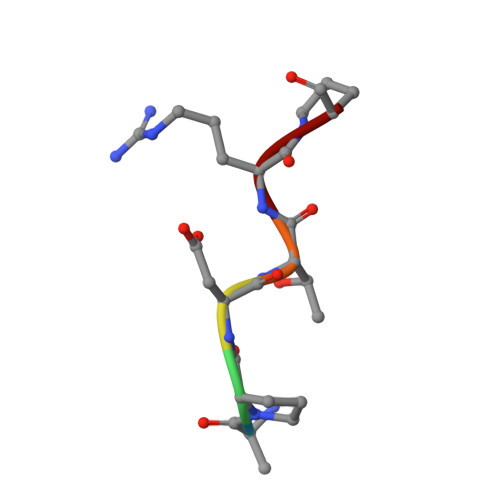
    The tumor-associated carbohydrate Tn antigens include two variants, αGalNAc- O-Thr and αGalNAc- O-Ser. In solution, they exhibit dissimilar shapes and dynamics and bind differently to the same protein receptor. Here, we demonstrate experimentally and theoretically that their conformational preferences in the gas phase are highly similar, revealing the essential role of water. We propose that water molecules prompt the rotation around the glycosidic linkage in the threonine derivative, shielding its hydrophobic methyl group and allowing an optimal solvation of the polar region of the antigen. The unusual arrangement of αGalNAc- O-Thr features a water molecule bound into a "pocket" between the sugar and the threonine. This mechanism is supported by trapping, for the first time, such localized water in the crystal structures of an antibody bound to two glycopeptides that comprise fluorinated Tn antigens in their structure. According to several reported X-ray structures, installing oxygenated amino acids in specific regions of the receptor capable of displacing the bridging water molecule to the bulk-solvent may facilitate the molecular recognition of the Tn antigen with threonine. Overall, our data also explain how water fine-tunes the 3D structure features of similar molecules, which in turn are behind their distinct biological activities.


  • Organizational Affiliation

    Departamento de Química, Centro de Investigación en Síntesis Química , Universidad de La Rioja , 26006 Logroño , Spain.


Macromolecules
Find similar proteins by:  (by identity cutoff)  |  3D Structure
Entity ID: 1
MoleculeChains Sequence LengthOrganismDetailsImage
scFv-1SM3,scFv-1SM3A [auth H]244Mus musculusMutation(s): 0 
Entity Groups  
Sequence Clusters30% Identity50% Identity70% Identity90% Identity95% Identity100% Identity
Sequence Annotations
Expand
  • Reference Sequence

Find similar proteins by:  Sequence   |   3D Structure  

Entity ID: 2
MoleculeChains Sequence LengthOrganismDetailsImage
Mucin-1B [auth P]6Homo sapiensMutation(s): 0 
UniProt & NIH Common Fund Data Resources
Find proteins for P15941 (Homo sapiens)
Explore P15941 
Go to UniProtKB:  P15941
PHAROS:  P15941
GTEx:  ENSG00000185499 
Entity Groups  
UniProt GroupP15941
Sequence Annotations
Expand
  • Reference Sequence
Small Molecules
Ligands 2 Unique
IDChains Name / Formula / InChI Key2D Diagram3D Interactions
EEN
Query on EEN

Download Ideal Coordinates CCD File 
M [auth P]2-deoxy-2-[(difluoroacetyl)amino]-alpha-D-galactopyranose
C8 H13 F2 N O6
QWUDJWPZSGMAGG-WWHASAIZSA-N
EDO
Query on EDO

Download Ideal Coordinates CCD File 
C [auth H]
D [auth H]
E [auth H]
F [auth H]
G [auth H]
C [auth H],
D [auth H],
E [auth H],
F [auth H],
G [auth H],
H,
I [auth H],
J [auth H],
K [auth H],
L [auth H]
1,2-ETHANEDIOL
C2 H6 O2
LYCAIKOWRPUZTN-UHFFFAOYSA-N
Experimental Data & Validation

Experimental Data

  • Method: X-RAY DIFFRACTION
  • Resolution: 1.70 Å
  • R-Value Free: 0.225 
  • R-Value Work: 0.175 
  • R-Value Observed: 0.177 
  • Space Group: P 21 21 21
Unit Cell:
Length ( Å )Angle ( ˚ )
a = 35.324α = 90
b = 69.639β = 90
c = 90.59γ = 90
Software Package:
Software NamePurpose
REFMACrefinement
XDSdata reduction
SCALAdata scaling
MOLREPphasing

Structure Validation

View Full Validation Report


Ligand Structure Quality Assessment 


Entry History & Funding Information

Deposition Data


Funding OrganizationLocationGrant Number
MICINNSpainCTQ2013-44367-C2-2-P
MICINNSpainBFU2016-75633-P

Revision History  (Full details and data files)

  • Version 1.0: 2019-02-20
    Type: Initial release
  • Version 1.1: 2019-09-04
    Changes: Data collection, Database references
  • Version 2.0: 2020-07-29
    Type: Remediation
    Reason: Carbohydrate remediation
    Changes: Atomic model, Derived calculations, Structure summary
  • Version 2.1: 2024-01-17
    Changes: Data collection, Database references, Refinement description, Structure summary