6FUI

Complement factor D in complex with the inhibitor 3-((3-((3-(aminomethyl)phenyl)amino)-1H-pyrazolo[3,4-d]pyrimidin-4-yl)amino)phenol


Experimental Data Snapshot

  • Method: X-RAY DIFFRACTION
  • Resolution: 1.38 Å
  • R-Value Free: 0.198 
  • R-Value Work: 0.132 
  • R-Value Observed: 0.136 

wwPDB Validation   3D Report Full Report


Ligand Structure Quality Assessment 


This is version 1.0 of the entry. See complete history


Literature

Discovery and Design of First Benzylamine-Based Ligands Binding to an Unlocked Conformation of the Complement Factor D.

Vulpetti, A.Ostermann, N.Randl, S.Yoon, T.Mac Sweeney, A.Cumin, F.Lorthiois, E.Rudisser, S.Erbel, P.Maibaum, J.

(2018) ACS Med Chem Lett 9: 490-495

  • DOI: https://doi.org/10.1021/acsmedchemlett.8b00104
  • Primary Citation of Related Structures:  
    6FTY, 6FTZ, 6FUG, 6FUH, 6FUI, 6FUJ, 6FUT

  • PubMed Abstract: 

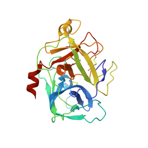
    Complement Factor D, a serine protease of the S1 family and key component of the alternative pathway amplification loop, represents a promising target for the treatment of several prevalent and rare diseases linked to the innate immune system. Previously reported FD inhibitors have been shown to bind to the FD active site in its self-inhibited conformation characterized by the presence of a salt bridge at the bottom of the S1 pocket between Asp189 and Arg218. We report herein a new set of small-molecule FD ligands that harbor a basic S1 binding moiety directly binding to the carboxylate of Asp189, thereby displacing the Asp189-Arg218 ionic interaction and significantly changing the conformation of the self-inhibitory loop.


  • Organizational Affiliation

    Novartis Pharma AG, Institutes for BioMedical Research, Novartis Campus, CH-4056 Basel, Switzerland.


Macromolecules
Find similar proteins by:  (by identity cutoff)  |  3D Structure
Entity ID: 1
MoleculeChains Sequence LengthOrganismDetailsImage
Complement factor D232Homo sapiensMutation(s): 0 
Gene Names: CFDDFPFD
EC: 3.4.21.46
UniProt & NIH Common Fund Data Resources
Find proteins for P00746 (Homo sapiens)
Explore P00746 
Go to UniProtKB:  P00746
PHAROS:  P00746
GTEx:  ENSG00000197766 
Entity Groups  
Sequence Clusters30% Identity50% Identity70% Identity90% Identity95% Identity100% Identity
UniProt GroupP00746
Sequence Annotations
Expand
  • Reference Sequence
Small Molecules
Ligands 1 Unique
IDChains Name / Formula / InChI Key2D Diagram3D Interactions
E7W (Subject of Investigation/LOI)
Query on E7W

Download Ideal Coordinates CCD File 
B [auth A](1~{R},2~{S})-2-[[4-[[3-(aminomethyl)phenyl]amino]quinazolin-2-yl]amino]cyclohexane-1-carboxylic acid
C22 H25 N5 O2
HGCXJWRMCKMKEG-MJGOQNOKSA-N
Binding Affinity Annotations 
IDSourceBinding Affinity
E7W BindingDB:  6FUI Kd: 3.50e+4 (nM) from 1 assay(s)
IC50: 8.40e+4 (nM) from 1 assay(s)
Binding MOAD:  6FUI Kd: 3.50e+4 (nM) from 1 assay(s)
Experimental Data & Validation

Experimental Data

  • Method: X-RAY DIFFRACTION
  • Resolution: 1.38 Å
  • R-Value Free: 0.198 
  • R-Value Work: 0.132 
  • R-Value Observed: 0.136 
  • Space Group: P 1 21 1
Unit Cell:
Length ( Å )Angle ( ˚ )
a = 55.508α = 90
b = 50.064β = 106.09
c = 39.264γ = 90
Software Package:
Software NamePurpose
REFMACrefinement
XDSdata reduction
XSCALEdata scaling
PHASERphasing

Structure Validation

View Full Validation Report


Ligand Structure Quality Assessment 


Entry History 

Deposition Data

Revision History  (Full details and data files)

  • Version 1.0: 2018-06-06
    Type: Initial release