


6F5M
Crystal structure of highly glycosylated human leukocyte elastase in complex with a thiazolidinedione inhibitor
- PDB DOI: https://doi.org/10.2210/pdb6F5M/pdb
- Classification: HYDROLASE
- Organism(s): Homo sapiens
- Mutation(s): No
- Deposited: 2017-12-01 Released: 2018-08-08
- Funding Organization(s): German Research Foundation
Experimental Data Snapshot
- Method: X-RAY DIFFRACTION
- Resolution: 2.70 Å
- R-Value Free: 0.234
- R-Value Work: 0.176
- R-Value Observed: 0.180

This is version 2.1 of the entry. See complete history.

Macromolecules
Find similar proteins by:
(by identity cutoff) | 3D Structure
Entity ID: 1 | |||||
|---|---|---|---|---|---|
| Molecule | Chains | Sequence Length | Organism | Details | Image |
| Neutrophil elastase | 218 | Homo sapiens | Mutation(s): 0 EC: 3.4.21.37 | 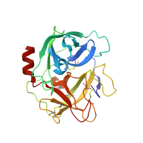 | |
UniProt & NIH Common Fund Data Resources | |||||
Find proteins for P08246 (Homo sapiens) Explore P08246 Go to UniProtKB: P08246 | |||||
PHAROS: P08246 GTEx: ENSG00000197561 | |||||
Entity Groups | |||||
| Sequence Clusters | 30% Identity50% Identity70% Identity90% Identity95% Identity100% Identity | ||||
| UniProt Group | P08246 | ||||
Sequence AnnotationsExpand | |||||
| |||||
Oligosaccharides
Entity ID: 2 | |||||
|---|---|---|---|---|---|
| Molecule | Chains | Length | 2D Diagram | Glycosylation | 3D Interactions |
| alpha-D-mannopyranose-(1-6)-beta-D-mannopyranose-(1-4)-2-acetamido-2-deoxy-beta-D-glucopyranose-(1-4)-[alpha-L-fucopyranose-(1-6)]2-acetamido-2-deoxy-beta-D-glucopyranose | C | 5 |  | N-Glycosylation | |
Glycosylation Resources | |||||
GlyTouCan: G00395TQ GlyCosmos: G00395TQ GlyGen: G00395TQ | |||||
Entity ID: 3 | |||||
|---|---|---|---|---|---|
| Molecule | Chains | Length | 2D Diagram | Glycosylation | 3D Interactions |
| beta-D-mannopyranose-(1-4)-2-acetamido-2-deoxy-beta-D-glucopyranose-(1-4)-[alpha-L-fucopyranose-(1-6)]2-acetamido-2-deoxy-beta-D-glucopyranose | D, F | 4 |  | N-Glycosylation | |
Glycosylation Resources | |||||
GlyTouCan: G32152BH GlyCosmos: G32152BH GlyGen: G32152BH | |||||
Entity ID: 4 | |||||
|---|---|---|---|---|---|
| Molecule | Chains | Length | 2D Diagram | Glycosylation | 3D Interactions |
| alpha-D-mannopyranose-(1-3)-[alpha-D-mannopyranose-(1-6)]beta-D-mannopyranose-(1-4)-2-acetamido-2-deoxy-beta-D-glucopyranose-(1-4)-[alpha-L-fucopyranose-(1-6)]2-acetamido-2-deoxy-beta-D-glucopyranose | E | 6 |  | N-Glycosylation | |
Glycosylation Resources | |||||
GlyTouCan: G82348BZ GlyCosmos: G82348BZ GlyGen: G82348BZ | |||||
Small Molecules
| Ligands 3 Unique | |||||
|---|---|---|---|---|---|
| ID | Chains | Name / Formula / InChI Key | 2D Diagram | 3D Interactions | |
| CQH Query on CQH | K [auth A], L [auth B] | 5-[[4-[[(2~{S})-4-methyl-1-oxidanylidene-1-[(2-propylphenyl)amino]pentan-2-yl]carbamoyl]phenyl]methyl]-2-oxidanylidene-1,3-thiazol-1-ium-4-olate C26 H29 N3 O4 S NWLPQCZZJXMUOM-NRFANRHFSA-N |  | ||
| SO4 Query on SO4 | G [auth A], H [auth A], M [auth B], N [auth B] | SULFATE ION O4 S QAOWNCQODCNURD-UHFFFAOYSA-L |  | ||
| ACT Query on ACT | I [auth A], J [auth A], O [auth B] | ACETATE ION C2 H3 O2 QTBSBXVTEAMEQO-UHFFFAOYSA-M |  | ||
Experimental Data & Validation
Experimental Data
- Method: X-RAY DIFFRACTION
- Resolution: 2.70 Å
- R-Value Free: 0.234
- R-Value Work: 0.176
- R-Value Observed: 0.180
- Space Group: H 3 2
Unit Cell:
| Length ( Å ) | Angle ( ˚ ) |
|---|---|
| a = 204.557 | α = 90 |
| b = 204.557 | β = 90 |
| c = 62.155 | γ = 120 |
| Software Name | Purpose |
|---|---|
| PHENIX | refinement |
| XDS | data reduction |
| Aimless | data scaling |
| PHASER | phasing |
Entry History & Funding Information
Deposition Data
- Released Date: 2018-08-08 Deposition Author(s): Hochscherf, J., Pietsch, M., Tieu, W., Kuan, K., Hautmann, S., Abell, A., Guetschow, M., Niefind, K.
| Funding Organization | Location | Grant Number |
|---|---|---|
| German Research Foundation | Germany | NI 643/4-1 |
Revision History (Full details and data files)
- Version 1.0: 2018-08-08
Type: Initial release - Version 1.1: 2018-08-15
Changes: Data collection, Database references - Version 2.0: 2020-07-29
Type: Remediation
Reason: Carbohydrate remediation
Changes: Atomic model, Data collection, Derived calculations, Structure summary - Version 2.1: 2024-01-17
Changes: Data collection, Database references, Derived calculations, Refinement description, Structure summary
