6EBX

STRUCTURE DETERMINATION OF A DIMERIC FORM OF ERABUTOXIN B, CRYSTALLIZED FROM THIOCYANATE SOLUTION


Experimental Data Snapshot

  • Method: X-RAY DIFFRACTION
  • Resolution: 1.70 Å
  • R-Value Work: 0.194 

wwPDB Validation   3D Report Full Report


This is version 1.3 of the entry. See complete history


Literature

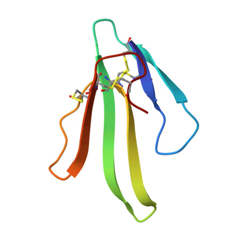
Structure determination of a dimeric form of erabutoxin-b, crystallized from a thiocyanate solution.

Saludjian, P.Prange, T.Navaza, J.Menez, R.Guilloteau, J.P.Ries-Kautt, M.Ducruix, A.

(1992) Acta Crystallogr B 48: 520-531

  • DOI: https://doi.org/10.1107/s010876819200096x
  • Primary Citation of Related Structures:  
    6EBX

  • PubMed Abstract: 

    Erabutoxin-b, M(r) = 6861.1, a single 62 amino-acid chain folded by four disulfide bridges, was crystallized in a new orthorhombic form by using thiocyanate as crystallizing agent. The space group is P2(1)2(1)2(1) with a = 53.36 (4), b = 40.89 (4), c = 55.71 (5) A, V = 121533.1 A and Z = 8. X-ray diffraction data were recorded at the LURE synchrotron facility (lambda = 1.405 A). The structure was solved by molecular replacement and shows a dimeric association through an anti-parallel beta-sheet around the twofold non-crystallographic axis. The two independent molecules, one SCN- ion and 97 associated water molecules were refined by molecular dynamics and annealing techniques to R = 19.6% (10,913 Fobs, resolution 5-1.7 A). The thiocyanate ion is located at the interface of the dimer and close to the non-crystallographic twofold axis.


  • Organizational Affiliation

    Chimie Structurale Bio-moléculaire (URA 1430 CNRS), UFR Biomédicale, Bobigny, France.


Macromolecules
Find similar proteins by:  (by identity cutoff)  |  3D Structure
Entity ID: 1
MoleculeChains Sequence LengthOrganismDetailsImage
ERABUTOXIN B
A, B
62Laticauda semifasciataMutation(s): 0 
UniProt
Find proteins for Q90VW1 (Laticauda semifasciata)
Explore Q90VW1 
Go to UniProtKB:  Q90VW1
Entity Groups  
Sequence Clusters30% Identity50% Identity70% Identity90% Identity95% Identity100% Identity
UniProt GroupQ90VW1
Sequence Annotations
Expand
  • Reference Sequence
Small Molecules
Ligands 1 Unique
IDChains Name / Formula / InChI Key2D Diagram3D Interactions
SCN
Query on SCN

Download Ideal Coordinates CCD File 
C [auth B]THIOCYANATE ION
C N S
ZMZDMBWJUHKJPS-UHFFFAOYSA-M
Experimental Data & Validation

Experimental Data

  • Method: X-RAY DIFFRACTION
  • Resolution: 1.70 Å
  • R-Value Work: 0.194 
  • Space Group: P 21 21 21
Unit Cell:
Length ( Å )Angle ( ˚ )
a = 53.36α = 90
b = 40.89β = 90
c = 55.71γ = 90
Software Package:
Software NamePurpose
X-PLORmodel building
X-PLORrefinement
X-PLORphasing

Structure Validation

View Full Validation Report


Entry History 

Deposition Data

Revision History  (Full details and data files)

  • Version 1.0: 1993-01-15
    Type: Initial release
  • Version 1.1: 2008-03-03
    Changes: Version format compliance
  • Version 1.2: 2011-07-13
    Changes: Version format compliance
  • Version 1.3: 2017-11-29
    Changes: Derived calculations, Other