6E66

Crystal structure of bacterial N-acetylglucosamine transferase NleB


Experimental Data Snapshot

  • Method: X-RAY DIFFRACTION
  • Resolution: 2.10 Å
  • R-Value Free: 0.241 
  • R-Value Work: 0.200 
  • R-Value Observed: 0.202 

wwPDB Validation   3D Report Full Report


This is version 1.1 of the entry. See complete history


Literature

Structural and Functional Insights into Host Death Domains Inactivation by the Bacterial Arginine GlcNAcyltransferase Effector.

Ding, J.Pan, X.Du, L.Yao, Q.Xue, J.Yao, H.Wang, D.C.Li, S.Shao, F.

(2019) Mol Cell 74: 922

  • DOI: https://doi.org/10.1016/j.molcel.2019.03.028
  • Primary Citation of Related Structures:  
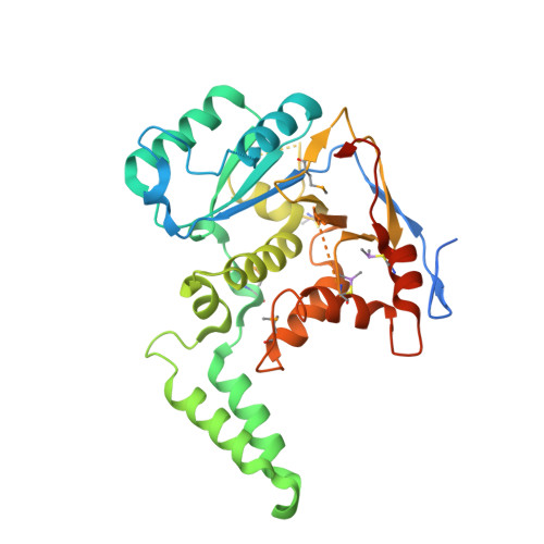
    6AC0, 6ACI, 6E66

  • PubMed Abstract: 

    Enteropathogenic E. coli NleB and related type III effectors catalyze arginine GlcNAcylation of death domain (DD) proteins to block host defense, but the underlying mechanism is unknown. Here we solve crystal structures of NleB alone and in complex with FADD-DD, UDP, and Mn 2+ as well as NleB-GlcNAcylated DDs of TRADD and RIPK1. NleB adopts a GT-A fold with a unique helix-pair insertion to hold FADD-DD; the interface contacts explain the selectivity of NleB for certain DDs. The acceptor arginine is fixed into a cleft, in which Glu253 serves as a base to activate the guanidinium. Analyses of the enzyme-substrate complex and the product structures reveal an inverting sugar-transfer reaction and a detailed catalytic mechanism. These structural insights are validated by mutagenesis analyses of NleB-mediated GlcNAcylation in vitro and its function in mouse infection. Our study builds a structural framework for understanding of NleB-catalyzed arginine GlcNAcylation of host death domain.


  • Organizational Affiliation

    National Laboratory of Biomacromolecules, CAS Center for Excellence in Biomacromolecules, Institute of Biophysics, Chinese Academy of Sciences, Beijing 100101, China; National Institute of Biological Sciences, Beijing 102206, China. Electronic address: jding@ibp.ac.cn.


Macromolecules
Find similar proteins by:  (by identity cutoff)  |  3D Structure
Entity ID: 1
MoleculeChains Sequence LengthOrganismDetailsImage
NleB330Escherichia coliMutation(s): 0 
UniProt
Find proteins for B7UI21 (Escherichia coli O127:H6 (strain E2348/69 / EPEC))
Explore B7UI21 
Go to UniProtKB:  B7UI21
Entity Groups  
Sequence Clusters30% Identity50% Identity70% Identity90% Identity95% Identity100% Identity
UniProt GroupB7UI21
Sequence Annotations
Expand
  • Reference Sequence
Small Molecules
Ligands 1 Unique
IDChains Name / Formula / InChI Key2D Diagram3D Interactions
EDO
Query on EDO

Download Ideal Coordinates CCD File 
B [auth A]1,2-ETHANEDIOL
C2 H6 O2
LYCAIKOWRPUZTN-UHFFFAOYSA-N
Modified Residues  2 Unique
IDChains TypeFormula2D DiagramParent
CAS
Query on CAS
A
L-PEPTIDE LINKINGC5 H12 As N O2 SCYS
MSE
Query on MSE
A
L-PEPTIDE LINKINGC5 H11 N O2 SeMET
Experimental Data & Validation

Experimental Data

  • Method: X-RAY DIFFRACTION
  • Resolution: 2.10 Å
  • R-Value Free: 0.241 
  • R-Value Work: 0.200 
  • R-Value Observed: 0.202 
  • Space Group: P 21 21 2
Unit Cell:
Length ( Å )Angle ( ˚ )
a = 85.587α = 90
b = 101.458β = 90
c = 38.636γ = 90
Software Package:
Software NamePurpose
PHENIXrefinement
HKL-2000data reduction
HKL-2000data scaling
PHENIXphasing

Structure Validation

View Full Validation Report


Entry History 

Deposition Data

Revision History  (Full details and data files)

  • Version 1.0: 2019-06-05
    Type: Initial release
  • Version 1.1: 2019-06-19
    Changes: Data collection, Database references