6DWI

Structure of the 4462 Antibody Fab fragment bound to a Staphylococcus aureus wall techoic acid analog


Experimental Data Snapshot

  • Method: X-RAY DIFFRACTION
  • Resolution: 2.39 Å
  • R-Value Free: 0.249 
  • R-Value Work: 0.203 
  • R-Value Observed: 0.206 

wwPDB Validation   3D Report Full Report


Ligand Structure Quality Assessment 


This is version 2.1 of the entry. See complete history


Literature

Structural investigation of human S. aureus-targeting antibodies that bind wall teichoic acid.

Fong, R.Kajihara, K.Chen, M.Hotzel, I.Mariathasan, S.Hazenbos, W.L.W.Lupardus, P.J.

(2018) MAbs 10: 979-991

  • DOI: https://doi.org/10.1080/19420862.2018.1501252
  • Primary Citation of Related Structures:  
    6DW2, 6DWA, 6DWC, 6DWI

  • PubMed Abstract: 

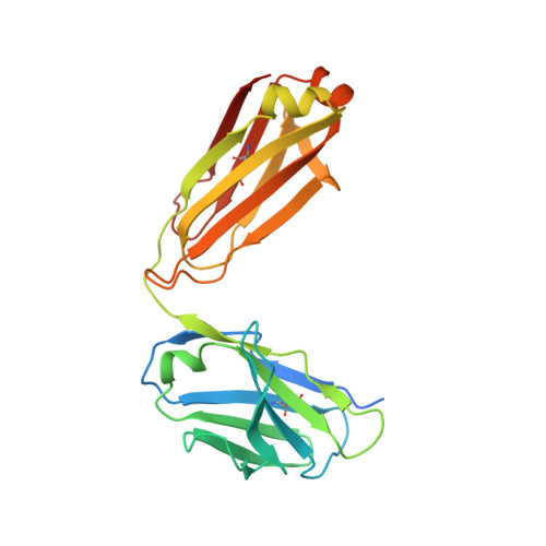
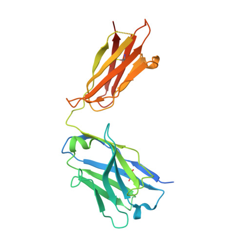
    Infections caused by methicillin-resistant Staphylococcus aureus (MRSA) are a growing health threat worldwide. Efforts to identify novel antibodies that target S. aureus cell surface antigens are a promising direction in the development of antibiotics that can halt MRSA infection. We biochemically and structurally characterized three patient-derived MRSA-targeting antibodies that bind to wall teichoic acid (WTA), which is a polyanionic surface glycopolymer. In S. aureus, WTA exists in both α- and β-forms, based on the stereochemistry of attachment of a N-acetylglucosamine residue to the repeating phosphoribitol sugar unit. We identified a panel of antibodies cloned from human patients that specifically recognize the α or β form of WTA, and can bind with high affinity to pathogenic wild-type strains of S. aureus bacteria. To investigate how the β-WTA specific antibodies interact with their target epitope, we determined the X-ray crystal structures of the three β-WTA specific antibodies, 4462, 4497, and 6078 (Protein Data Bank IDs 6DWI, 6DWA, and 6DW2, respectively), bound to a synthetic WTA epitope. These structures reveal that all three of these antibodies, while utilizing distinct antibody complementarity-determining region sequences and conformations to interact with β-WTA, fulfill two recognition principles: binding to the β-GlcNAc pyranose core and triangulation of WTA phosphate residues with polar contacts. These studies reveal the molecular basis for targeting a unique S. aureus cell surface epitope and highlight the power of human patient-based antibody discovery techniques for finding novel pathogen-targeting therapeutics.


  • Organizational Affiliation

    a Department of Structural Biology , Genentech , South San Francisco , CA , USA.


Macromolecules
Find similar proteins by:  (by identity cutoff)  |  3D Structure
Entity ID: 1
MoleculeChains Sequence LengthOrganismDetailsImage
4462 Fab Light Chain
A, C, E, G, I
A, C, E, G, I, K, M, O
233Homo sapiensMutation(s): 0 
Entity Groups  
Sequence Clusters30% Identity50% Identity70% Identity90% Identity95% Identity100% Identity
Sequence Annotations
Expand
  • Reference Sequence
Find similar proteins by:  (by identity cutoff)  |  3D Structure
Entity ID: 2
MoleculeChains Sequence LengthOrganismDetailsImage
4462 Fab Heavy chain
B, D, F, H, J
B, D, F, H, J, L, N, P
243Homo sapiensMutation(s): 0 
Entity Groups  
Sequence Clusters30% Identity50% Identity70% Identity90% Identity95% Identity100% Identity
Sequence Annotations
Expand
  • Reference Sequence
Small Molecules
Ligands 4 Unique
IDChains Name / Formula / InChI Key2D Diagram3D Interactions
HD4
Query on HD4

Download Ideal Coordinates CCD File 
BA [auth L],
T [auth D],
X [auth H],
Z [auth J]
4-O-[2-acetamido-2-deoxy-beta-D-glucopyranosyl]-1-O-phosphono-D-ribitol
C13 H26 N O13 P
UTDJSTSCOBYMHN-SKCKNYEVSA-N
FLC
Query on FLC

Download Ideal Coordinates CCD File 
W [auth G]CITRATE ANION
C6 H5 O7
KRKNYBCHXYNGOX-UHFFFAOYSA-K
GOL
Query on GOL

Download Ideal Coordinates CCD File 
AA [auth J],
Y [auth H]
GLYCEROL
C3 H8 O3
PEDCQBHIVMGVHV-UHFFFAOYSA-N
CL
Query on CL

Download Ideal Coordinates CCD File 
CA [auth O]
Q [auth A]
R [auth B]
S [auth C]
U [auth F]
CA [auth O],
Q [auth A],
R [auth B],
S [auth C],
U [auth F],
V [auth G]
CHLORIDE ION
Cl
VEXZGXHMUGYJMC-UHFFFAOYSA-M
Experimental Data & Validation

Experimental Data

  • Method: X-RAY DIFFRACTION
  • Resolution: 2.39 Å
  • R-Value Free: 0.249 
  • R-Value Work: 0.203 
  • R-Value Observed: 0.206 
  • Space Group: P 1 21 1
Unit Cell:
Length ( Å )Angle ( ˚ )
a = 122.573α = 90
b = 114.097β = 90.46
c = 128.301γ = 90
Software Package:
Software NamePurpose
BUSTERrefinement
HKL-2000data reduction
SCALEPACKdata scaling
PHASERphasing

Structure Validation

View Full Validation Report


Ligand Structure Quality Assessment 


Entry History 

Deposition Data

Revision History  (Full details and data files)

  • Version 1.0: 2018-08-29
    Type: Initial release
  • Version 1.1: 2018-09-05
    Changes: Data collection, Database references
  • Version 1.2: 2018-10-10
    Changes: Data collection, Database references
  • Version 2.0: 2020-07-29
    Type: Remediation
    Reason: Carbohydrate remediation
    Changes: Atomic model, Derived calculations, Structure summary
  • Version 2.1: 2023-10-11
    Changes: Advisory, Data collection, Database references, Refinement description, Structure summary