6DUM

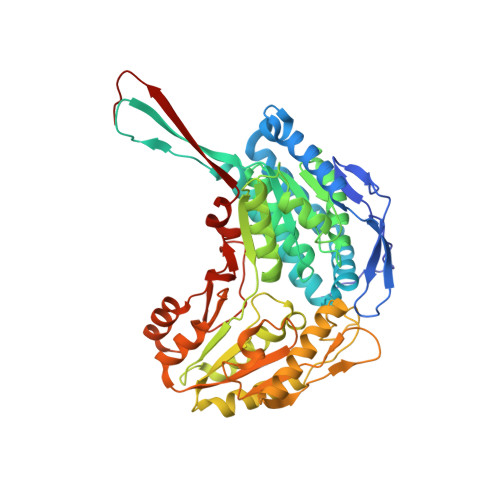
ALDH1A1 N121S in complex with 6-{[(3-fluorophenyl)methyl]sulfanyl}-2-(oxetan-3-yl)-5-phenyl-2,5-dihydro-4H-pyrazolo[3,4-d]pyrimidin-4-one (compound 13g)


Experimental Data Snapshot

  • Method: X-RAY DIFFRACTION
  • Resolution: 2.00 Å
  • R-Value Free: 0.249 
  • R-Value Work: 0.194 
  • R-Value Observed: 0.197 

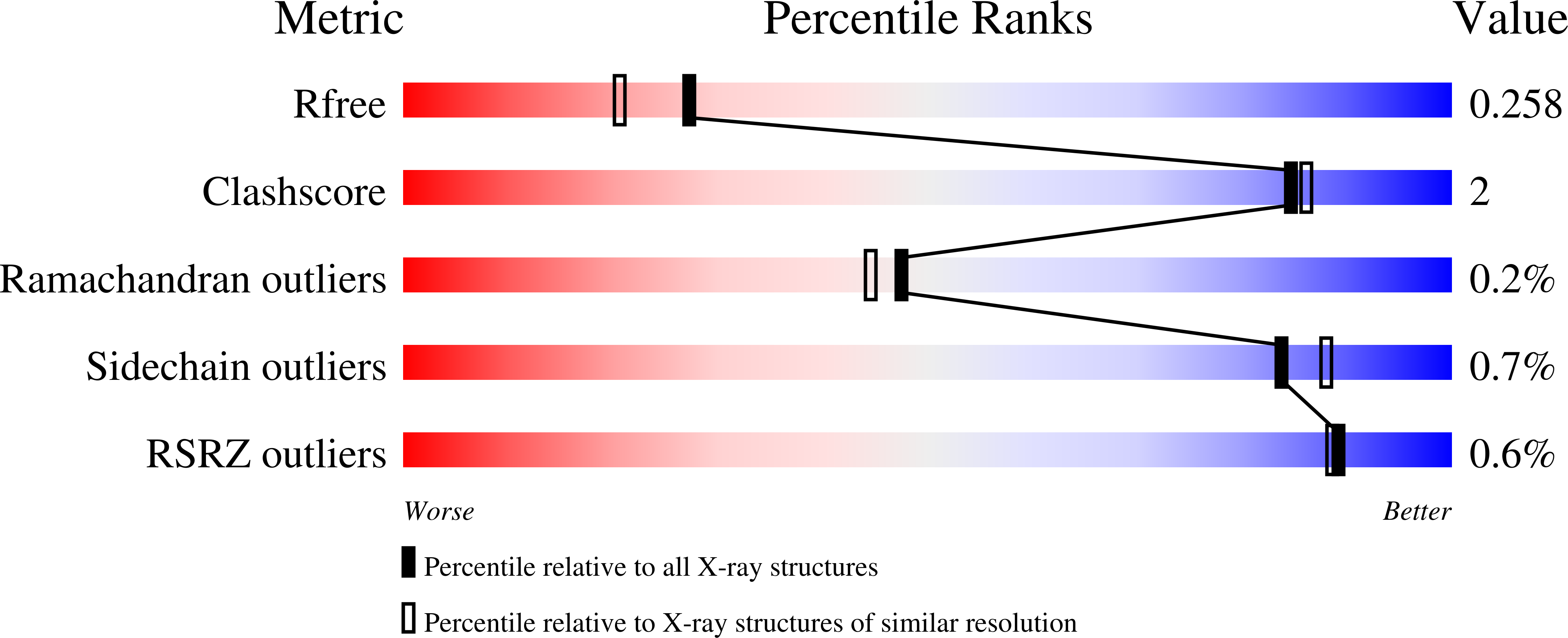
wwPDB Validation   3D Report Full Report


Ligand Structure Quality Assessment 


This is version 1.2 of the entry. See complete history


Literature

Structure-Based Optimization of a Novel Class of Aldehyde Dehydrogenase 1A (ALDH1A) Subfamily-Selective Inhibitors as Potential Adjuncts to Ovarian Cancer Chemotherapy.

Huddle, B.C.Grimley, E.Buchman, C.D.Chtcherbinine, M.Debnath, B.Mehta, P.Yang, K.Morgan, C.A.Li, S.Felton, J.Sun, D.Mehta, G.Neamati, N.Buckanovich, R.J.Hurley, T.D.Larsen, S.D.

(2018) J Med Chem 61: 8754-8773

  • DOI: https://doi.org/10.1021/acs.jmedchem.8b00930
  • Primary Citation of Related Structures:  
    6DUM

  • PubMed Abstract: 

    Aldehyde dehydrogenase (ALDH) activity is commonly used as a marker to identify cancer stem-like cells. The three ALDH1A isoforms have all been individually implicated in cancer stem-like cells and in chemoresistance; however, which isoform is preferentially expressed varies between cell lines. We sought to explore the structural determinants of ALDH1A isoform selectivity in a series of small-molecule inhibitors in support of research into the role of ALDH1A in cancer stem cells. An SAR campaign guided by a cocrystal structure of the HTS hit CM39 (7) with ALDH1A1 afforded first-in-class inhibitors of the ALDH1A subfamily with excellent selectivity over the homologous ALDH2 isoform. We also discovered the first reported modestly selective single isoform 1A2 and 1A3 inhibitors. Two compounds, 13g and 13h, depleted the CD133 + putative cancer stem cell pool, synergized with cisplatin, and achieved efficacious concentrations in vivo following IP administration. Compound 13h additionally synergized with cisplatin in a patient-derived ovarian cancer spheroid model.


  • Organizational Affiliation

    Department of Biochemistry and Molecular Biology , Indiana University School of Medicine , Indianapolis , Indiana 46202 , United States.


Macromolecules
Find similar proteins by:  (by identity cutoff)  |  3D Structure
Entity ID: 1
MoleculeChains Sequence LengthOrganismDetailsImage
Retinal dehydrogenase 1501Homo sapiensMutation(s): 0 
Gene Names: ALDH1A1ALDCALDH1PUMB1
EC: 1.2.1 (PDB Primary Data), 1.2.1.36 (PDB Primary Data)
UniProt & NIH Common Fund Data Resources
Find proteins for P00352 (Homo sapiens)
Explore P00352 
Go to UniProtKB:  P00352
PHAROS:  P00352
GTEx:  ENSG00000165092 
Entity Groups  
Sequence Clusters30% Identity50% Identity70% Identity90% Identity95% Identity100% Identity
UniProt GroupP00352
Sequence Annotations
Expand
  • Reference Sequence
Small Molecules
Ligands 4 Unique
IDChains Name / Formula / InChI Key2D Diagram3D Interactions
NAI
Query on NAI

Download Ideal Coordinates CCD File 
E [auth A]1,4-DIHYDRONICOTINAMIDE ADENINE DINUCLEOTIDE
C21 H29 N7 O14 P2
BOPGDPNILDQYTO-NNYOXOHSSA-N
A5Y
Query on A5Y

Download Ideal Coordinates CCD File 
D [auth A]6-{[(3-fluorophenyl)methyl]sulfanyl}-2-(oxetan-3-yl)-5-phenyl-2,5-dihydro-4H-pyrazolo[3,4-d]pyrimidin-4-one
C21 H17 F N4 O2 S
IBYYWHNJSYBLSJ-UHFFFAOYSA-N
YB
Query on YB

Download Ideal Coordinates CCD File 
B [auth A],
C [auth A]
YTTERBIUM (III) ION
Yb
AWSFICBXMUKWSK-UHFFFAOYSA-N
CL
Query on CL

Download Ideal Coordinates CCD File 
F [auth A]
G [auth A]
H [auth A]
I [auth A]
J [auth A]
F [auth A],
G [auth A],
H [auth A],
I [auth A],
J [auth A],
K [auth A],
L [auth A]
CHLORIDE ION
Cl
VEXZGXHMUGYJMC-UHFFFAOYSA-M
Binding Affinity Annotations 
IDSourceBinding Affinity
A5Y BindingDB:  6DUM IC50: 80 (nM) from 1 assay(s)
Binding MOAD:  6DUM IC50: 80 (nM) from 1 assay(s)
Experimental Data & Validation

Experimental Data

  • Method: X-RAY DIFFRACTION
  • Resolution: 2.00 Å
  • R-Value Free: 0.249 
  • R-Value Work: 0.194 
  • R-Value Observed: 0.197 
  • Space Group: P 4 2 2
Unit Cell:
Length ( Å )Angle ( ˚ )
a = 108.802α = 90
b = 108.802β = 90
c = 82.877γ = 90
Software Package:
Software NamePurpose
REFMACrefinement
SCALEPACKdata scaling
PDB_EXTRACTdata extraction

Structure Validation

View Full Validation Report


Ligand Structure Quality Assessment 


Entry History & Funding Information

Deposition Data


Funding OrganizationLocationGrant Number
National Institutes of Health/National Cancer Institute (NIH/NCI)United States--

Revision History  (Full details and data files)

  • Version 1.0: 2019-05-01
    Type: Initial release
  • Version 1.1: 2019-12-04
    Changes: Author supporting evidence
  • Version 1.2: 2024-03-13
    Changes: Data collection, Database references, Derived calculations