6DK4

Crystal structure of Campylobacter jejuni peroxide stress regulator


Experimental Data Snapshot

  • Method: X-RAY DIFFRACTION
  • Resolution: 2.71 Å
  • R-Value Free: 0.270 
  • R-Value Work: 0.215 
  • R-Value Observed: 0.218 

wwPDB Validation   3D Report Full Report


This is version 1.3 of the entry. See complete history


Literature

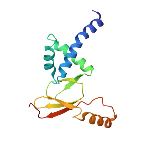
Crystal structure of Campylobacter jejuni peroxide regulator.

Sarvan, S.Charih, F.Butcher, J.Brunzelle, J.S.Stintzi, A.Couture, J.F.

(2018) FEBS Lett 592: 2351-2360

  • DOI: https://doi.org/10.1002/1873-3468.13120
  • Primary Citation of Related Structures:  
    6DK4

  • PubMed Abstract: 

    In Campylobacter jejuni (Cj), the metal-cofactored peroxide response regulator (PerR) transcription factor allows C. jejuni to respond to oxidative stresses. The crystal structure of the metalated form of CjPerR shows that the protein folds as an asymmetric dimer displaying structural differences in the orientation of its DNA-binding domain. Comparative analysis shows that such asymmetry is a conserved feature among crystallized PerR proteins, and mutational analysis reveals that residues found in the first α-helix of CjPerR contribute to DNA binding. These studies present the structure of CjPerR protein and highlight structural heterogeneity in the orientation of the metalated PerR DNA-binding domain which may underlie the ability of PerR to recognize DNA, control gene expression, and contribute to bacterial pathogenesis.


  • Organizational Affiliation

    Department of Biochemistry, Microbiology and Immunology, Ottawa Institute of Systems Biology, University of Ottawa, Canada.


Macromolecules
Find similar proteins by:  (by identity cutoff)  |  3D Structure
Entity ID: 1
MoleculeChains Sequence LengthOrganismDetailsImage
Ferric uptake regulation protein
A, B
142Campylobacter jejuniMutation(s): 0 
Gene Names: perRfurCj0322
UniProt
Find proteins for Q0PBI7 (Campylobacter jejuni subsp. jejuni serotype O:2 (strain ATCC 700819 / NCTC 11168))
Explore Q0PBI7 
Go to UniProtKB:  Q0PBI7
Entity Groups  
Sequence Clusters30% Identity50% Identity70% Identity90% Identity95% Identity100% Identity
UniProt GroupQ0PBI7
Sequence Annotations
Expand
  • Reference Sequence
Small Molecules
Experimental Data & Validation

Experimental Data

  • Method: X-RAY DIFFRACTION
  • Resolution: 2.71 Å
  • R-Value Free: 0.270 
  • R-Value Work: 0.215 
  • R-Value Observed: 0.218 
  • Space Group: P 21 21 21
Unit Cell:
Length ( Å )Angle ( ˚ )
a = 48.11α = 90
b = 70.186β = 90
c = 85.158γ = 90
Software Package:
Software NamePurpose
BUSTERrefinement
XDSdata reduction
autoPROCdata scaling
AutoSolphasing

Structure Validation

View Full Validation Report


Entry History & Funding Information

Deposition Data


Funding OrganizationLocationGrant Number
Canadian Institutes of Health Research (CIHR)Canada--

Revision History  (Full details and data files)

  • Version 1.0: 2018-06-13
    Type: Initial release
  • Version 1.1: 2018-09-12
    Changes: Data collection, Database references
  • Version 1.2: 2020-01-08
    Changes: Author supporting evidence
  • Version 1.3: 2024-03-06
    Changes: Data collection, Database references, Refinement description