6DJ2

HIV-1 protease with single mutation L76V in complex with Lopinavir


Experimental Data Snapshot

  • Method: X-RAY DIFFRACTION
  • Resolution: 1.36 Å
  • R-Value Free: 0.196 
  • R-Value Observed: 0.148 

wwPDB Validation   3D Report Full Report


Ligand Structure Quality Assessment 


This is version 1.2 of the entry. See complete history


Literature

Drug Resistance Mutation L76V Alters Nonpolar Interactions at the Flap-Core Interface of HIV-1 Protease.

Wong-Sam, A.Wang, Y.F.Zhang, Y.Ghosh, A.K.Harrison, R.W.Weber, I.T.

(2018) ACS Omega 3: 12132-12140

  • DOI: https://doi.org/10.1021/acsomega.8b01683
  • Primary Citation of Related Structures:  
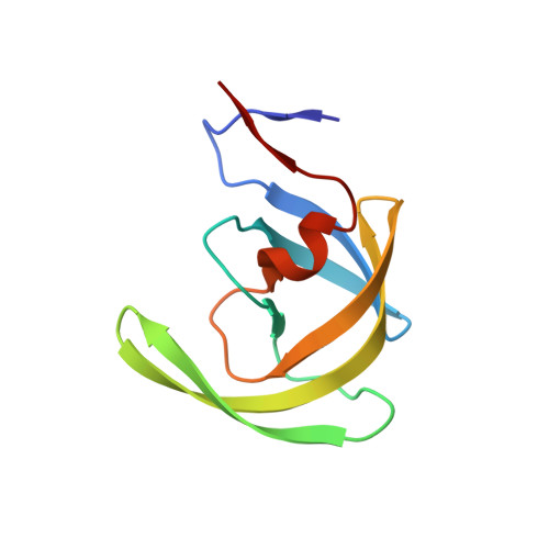
    6DIF, 6DIL, 6DJ1, 6DJ2, 6DJ5, 6DJ7

  • PubMed Abstract: 

    Four HIV-1 protease (PR) inhibitors, clinical inhibitors lopinavir and tipranavir, and two investigational compounds 4 and 5 , were studied for their effect on the structure and activity of PR with drug-resistant mutation L76V (PR L76V ). Compound 5 exhibited the best K i value of 1.9 nM for PR L76V , whereas the other three inhibitors had K i values of 4.5-7.6 nM, 2-3 orders of magnitude worse than for wild-type enzymes. Crystal structures showed only minor differences in interactions of inhibitors with PR L76V compared to wild-type complexes. The shorter side chain of Val76 in the mutant lost hydrophobic interactions with Lys45 and Ile47 in the flap, and with Asp30 and Thr74 in the protein core, consistent with decreased stability. Inhibitors forming additional polar interactions with the flaps or dimer interface of PR L76V were unable to compensate for the decrease in internal hydrophobic contacts. These structures provide insights for inhibitor design.


  • Organizational Affiliation

    Department of Biology, Molecular Basis of Disease Program, Department of Computer Science, and Department of Chemistry, Georgia State University, Atlanta, Georgia 30303, United States.


Macromolecules
Find similar proteins by:  (by identity cutoff)  |  3D Structure
Entity ID: 1
MoleculeChains Sequence LengthOrganismDetailsImage
HIV-1 protease
A, B
99Human immunodeficiency virus 1Mutation(s): 6 
Gene Names: pol
UniProt
Find proteins for P04585 (Human immunodeficiency virus type 1 group M subtype B (isolate HXB2))
Explore P04585 
Go to UniProtKB:  P04585
Entity Groups  
Sequence Clusters30% Identity50% Identity70% Identity90% Identity95% Identity100% Identity
UniProt GroupP04585
Sequence Annotations
Expand
  • Reference Sequence
Small Molecules
Binding Affinity Annotations 
IDSourceBinding Affinity
AB1 BindingDB:  6DJ2 Ki: min: 5.00e-3, max: 16 (nM) from 12 assay(s)
Kd: 1.30e-3 (nM) from 1 assay(s)
IC50: min: 2.5, max: 25 (nM) from 3 assay(s)
EC50: 1160 (nM) from 1 assay(s)
Binding MOAD:  6DJ2 Ki: 4.5 (nM) from 1 assay(s)
Experimental Data & Validation

Experimental Data

  • Method: X-RAY DIFFRACTION
  • Resolution: 1.36 Å
  • R-Value Free: 0.196 
  • R-Value Observed: 0.148 
  • Space Group: P 21 21 2
Unit Cell:
Length ( Å )Angle ( ˚ )
a = 60.011α = 90
b = 85.377β = 90
c = 46.43γ = 90
Software Package:
Software NamePurpose
HKL-2000data reduction
HKL-2000data scaling
PHASERphasing
SHELXL-97refinement

Structure Validation

View Full Validation Report


Ligand Structure Quality Assessment 


Entry History & Funding Information

Deposition Data


Funding OrganizationLocationGrant Number
National Institutes of Health/National Human Genome Research Institute (NIH/NHGRI)United StatesGM062920

Revision History  (Full details and data files)

  • Version 1.0: 2018-10-17
    Type: Initial release
  • Version 1.1: 2019-12-18
    Changes: Author supporting evidence
  • Version 1.2: 2023-10-11
    Changes: Data collection, Database references, Derived calculations, Refinement description, Structure summary