6DC7

Apo Fab structure of mouse monoclonal antibody 8B2

  • Classification: IMMUNE SYSTEM
  • Organism(s): Mus musculus
  • Mutation(s): No 

  • Deposited: 2018-05-04 Released: 2019-03-20 
  • Deposition Author(s): Chukwu, J.E., Kong, X.-P.
  • Funding Organization(s): National Institutes of Health/National Institute of Neurological Disorders and Stroke (NIH/NINDS), National Institutes of Health/National Institute on Aging (NIH/NIA)

Experimental Data Snapshot

  • Method: X-RAY DIFFRACTION
  • Resolution: 1.90 Å
  • R-Value Free: 0.235 
  • R-Value Work: 0.183 
  • R-Value Observed: 0.185 

wwPDB Validation   3D Report Full Report


This is version 1.2 of the entry. See complete history


Literature

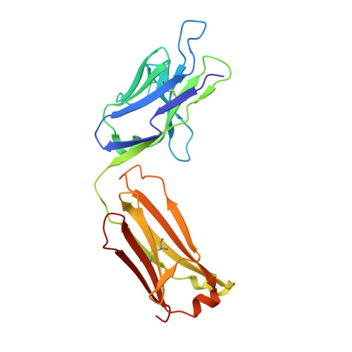
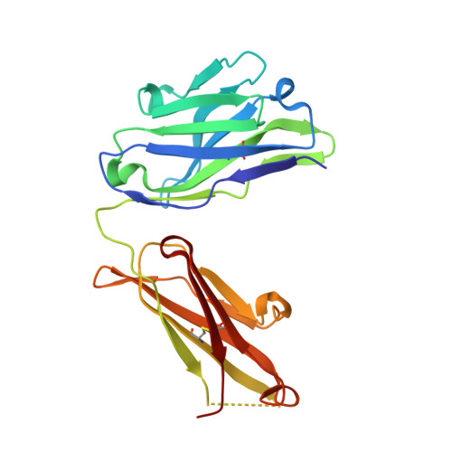
Structural characterization of monoclonal antibodies targeting C-terminal Ser404region of phosphorylated tau protein.

Chukwu, J.E.Congdon, E.E.Sigurdsson, E.M.Kong, X.P.

(2019) MAbs 11: 477-488

  • DOI: https://doi.org/10.1080/19420862.2019.1574530
  • Primary Citation of Related Structures:  
    6DC7, 6DC8, 6DC9, 6DCA

  • PubMed Abstract: 

    Targeting tau with immunotherapies is currently the most common approach taken in clinical trials of patients with Alzheimer's disease. The most prominent pathological feature of tau is its hyperphosphorylation, which may cause the protein to aggregate into toxic assemblies that collectively lead to neurodegeneration. Of the phospho-epitopes, the region around Ser 396 /Ser 404 has received particular attention for therapeutic targeting because of its prominence and stability in diseased tissue. Herein, we present the antigen-binding fragment (Fab)/epitope complex structures of three different monoclonal antibodies (mAbs) that target the pSer 404 tau epitope region. Most notably, these structures reveal an antigen conformation similar to a previously described pathogenic tau epitope, pSer 422 , which was shown to have a β-strand structure that may be linked to the seeding core in tau oligomers. In addition, we have previously reported on the similarly ordered conformation observed in a pSer 396 epitope, which is in tandem with pSer 404 . Our data are the first Fab structures of mAbs bound to this epitope region of the tau protein and support the existence of proteopathic tau conformations stabilized by specific phosphorylation events that are viable targets for immune modulation.


  • Organizational Affiliation

    a Department of Biochemistry & Molecular Pharmacology , New York University School of Medicine , New York , NY , USA.


Macromolecules
Find similar proteins by:  (by identity cutoff)  |  3D Structure
Entity ID: 1
MoleculeChains Sequence LengthOrganismDetailsImage
Fab light chainA [auth L],
C [auth M]
218Mus musculusMutation(s): 0 
Entity Groups  
Sequence Clusters30% Identity50% Identity70% Identity90% Identity95% Identity100% Identity
Sequence Annotations
Expand
  • Reference Sequence
Find similar proteins by:  (by identity cutoff)  |  3D Structure
Entity ID: 2
MoleculeChains Sequence LengthOrganismDetailsImage
Fab heavy chainB [auth H],
D [auth I]
217Mus musculusMutation(s): 0 
Entity Groups  
Sequence Clusters30% Identity50% Identity70% Identity90% Identity95% Identity100% Identity
Sequence Annotations
Expand
  • Reference Sequence
Experimental Data & Validation

Experimental Data

  • Method: X-RAY DIFFRACTION
  • Resolution: 1.90 Å
  • R-Value Free: 0.235 
  • R-Value Work: 0.183 
  • R-Value Observed: 0.185 
  • Space Group: P 2 21 21
Unit Cell:
Length ( Å )Angle ( ˚ )
a = 53.44α = 90
b = 67.474β = 90
c = 240.163γ = 90
Software Package:
Software NamePurpose
PHENIXrefinement
XDSdata reduction
XDSdata scaling
PHASERphasing
Cootmodel building

Structure Validation

View Full Validation Report


Entry History & Funding Information

Deposition Data


Funding OrganizationLocationGrant Number
National Institutes of Health/National Institute of Neurological Disorders and Stroke (NIH/NINDS)United StatesNS077239
National Institutes of Health/National Institute on Aging (NIH/NIA)United StatesAG032611

Revision History  (Full details and data files)

  • Version 1.0: 2019-03-20
    Type: Initial release
  • Version 1.1: 2019-04-03
    Changes: Data collection, Database references
  • Version 1.2: 2019-12-18
    Changes: Author supporting evidence