6CHA

STRUCTURE OF A TETRAHEDRAL TRANSITION STATE COMPLEX OF ALPHA-*CHYMOTRYPSIN AT 1.8-*ANGSTROMS RESOLUTION


Experimental Data Snapshot

  • Method: X-RAY DIFFRACTION
  • Resolution: 1.80 Å
  • R-Value Observed: 0.200 

wwPDB Validation   3D Report Full Report


This is version 1.2 of the entry. See complete history


Literature

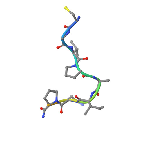
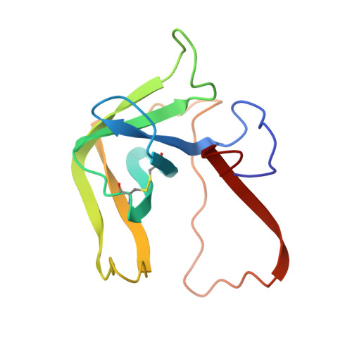
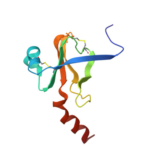
Structure of a tetrahedral transition state complex of alpha-chymotrypsin dimer at 1.8-A resolution.

Tulinsky, A.Blevins, R.A.

(1987) J Biol Chem 262: 7737-7743

  • DOI: https://doi.org/10.2210/pdb6cha/pdb
  • Primary Citation of Related Structures:  
    6CHA

  • PubMed Abstract: 

    A 1.8-A resolution x-ray crystallographic restrained least squares refinement has been carried out on the phenylethane boronic acid (PEBA) complex of alpha-chymotrypsin dimer (alpha-CHT), and it has been compared to the 1.67-A resolution structure of the native enzyme. PEBA has a high binding affinity for alpha-CHT, and the boronate forms a tetrahedral complex with Ser-195 OG of one molecule of the dimer; the boronate in the other molecule is severely disordered and does not form a tetrahedral complex. The former could be a model of the transition state of catalysis. The complex of PEBA X alpha-CHT displays significant nonequivalence in conformation of side chains between the independent molecules comparable to the native enzyme, but, like the latter, shows a high degree of fidelity in the folding of the main chain. The orientation of the phenyl ring, CA and CB of PEBA, in the specificity sites of the two molecules is similar, suggesting that recognition is fairly insensitive to small departures from local symmetry; the same does not apply to the boronate functionalities suggesting that greater precision is required for catalysis. The folding of the molecule remains the same upon PEBA binding, but some of the side chains respond nonequivalently. The latter is a consequence of the inherent nonequivalence of the native dimer and the asymmetrical nature of the PEBA binding.


Macromolecules

Find similar proteins by:  Sequence   |   3D Structure  

Entity ID: 1
MoleculeChains Sequence LengthOrganismDetailsImage
ALPHA-CHYMOTRYPSIN AA,
D [auth E]
13Bos taurusMutation(s): 0 
EC: 3.4.21.1
UniProt
Find proteins for P00766 (Bos taurus)
Explore P00766 
Go to UniProtKB:  P00766
Entity Groups  
Sequence Clusters30% Identity50% Identity70% Identity90% Identity95% Identity100% Identity
UniProt GroupP00766
Sequence Annotations
Expand
  • Reference Sequence
Find similar proteins by:  (by identity cutoff)  |  3D Structure
Entity ID: 2
MoleculeChains Sequence LengthOrganismDetailsImage
ALPHA-CHYMOTRYPSIN AB,
E [auth F]
131Bos taurusMutation(s): 0 
EC: 3.4.21.1
UniProt
Find proteins for P00766 (Bos taurus)
Explore P00766 
Go to UniProtKB:  P00766
Entity Groups  
Sequence Clusters30% Identity50% Identity70% Identity90% Identity95% Identity100% Identity
UniProt GroupP00766
Sequence Annotations
Expand
  • Reference Sequence
Find similar proteins by:  (by identity cutoff)  |  3D Structure
Entity ID: 3
MoleculeChains Sequence LengthOrganismDetailsImage
ALPHA-CHYMOTRYPSIN AC,
F [auth G]
97Bos taurusMutation(s): 0 
EC: 3.4.21.1
UniProt
Find proteins for P00766 (Bos taurus)
Explore P00766 
Go to UniProtKB:  P00766
Entity Groups  
Sequence Clusters30% Identity50% Identity70% Identity90% Identity95% Identity100% Identity
UniProt GroupP00766
Sequence Annotations
Expand
  • Reference Sequence
Small Molecules
Ligands 1 Unique
IDChains Name / Formula / InChI Key2D Diagram3D Interactions
PBA
Query on PBA

Download Ideal Coordinates CCD File 
G [auth C],
H [auth G]
PHENYLETHANE BORONIC ACID
C8 H11 B O2
VPRUMANMDWQMNF-UHFFFAOYSA-N
Binding Affinity Annotations 
IDSourceBinding Affinity
PBA Binding MOAD:  6CHA Ki: 4.00e+4 (nM) from 1 assay(s)
BindingDB:  6CHA Ki: 40 (nM) from 1 assay(s)
PDBBind:  6CHA Ki: 4.00e+4 (nM) from 1 assay(s)
Experimental Data & Validation

Experimental Data

  • Method: X-RAY DIFFRACTION
  • Resolution: 1.80 Å
  • R-Value Observed: 0.200 
  • Space Group: P 1 21 1
Unit Cell:
Length ( Å )Angle ( ˚ )
a = 49.27α = 90
b = 67.16β = 101.68
c = 65.91γ = 90
Software Package:
Software NamePurpose
PROLSQrefinement

Structure Validation

View Full Validation Report


Entry History 

Deposition Data

Revision History  (Full details and data files)

  • Version 1.0: 1987-04-16
    Type: Initial release
  • Version 1.1: 2008-03-03
    Changes: Version format compliance
  • Version 1.2: 2011-07-13
    Changes: Version format compliance