6CC4

Structure of MurJ from Escherichia coli


Experimental Data Snapshot

  • Method: X-RAY DIFFRACTION
  • Resolution: 3.50 Å
  • R-Value Free: 0.301 
  • R-Value Work: 0.280 
  • R-Value Observed: 0.281 

wwPDB Validation   3D Report Full Report


This is version 1.3 of the entry. See complete history


Literature

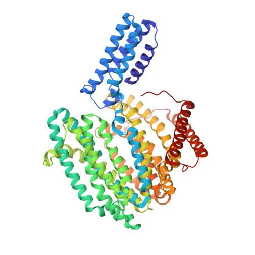
Structure and mutagenic analysis of the lipid II flippase MurJ fromEscherichia coli.

Zheng, S.Sham, L.T.Rubino, F.A.Brock, K.P.Robins, W.P.Mekalanos, J.J.Marks, D.S.Bernhardt, T.G.Kruse, A.C.

(2018) Proc Natl Acad Sci U S A 115: 6709-6714

  • DOI: https://doi.org/10.1073/pnas.1802192115
  • Primary Citation of Related Structures:  
    6CC4

  • PubMed Abstract: 

    The peptidoglycan cell wall provides an essential protective barrier in almost all bacteria, defining cellular morphology and conferring resistance to osmotic stress and other environmental hazards. The precursor to peptidoglycan, lipid II, is assembled on the inner leaflet of the plasma membrane. However, peptidoglycan polymerization occurs on the outer face of the plasma membrane, and lipid II must be flipped across the membrane by the MurJ protein before its use in peptidoglycan synthesis. Due to its central role in cell wall assembly, MurJ is of fundamental importance in microbial cell biology and is a prime target for novel antibiotic development. However, relatively little is known regarding the mechanisms of MurJ function, and structural data for MurJ are available only from the extremophile Thermosipho africanus Here, we report the crystal structure of substrate-free MurJ from the gram-negative model organism Escherichia coli , revealing an inward-open conformation. Taking advantage of the genetic tractability of E. coli , we performed high-throughput mutagenesis and next-generation sequencing to assess mutational tolerance at every amino acid in the protein, providing a detailed functional and structural map for the enzyme and identifying sites for inhibitor development. Lastly, through the use of sequence coevolution analysis, we identify functionally important interactions in the outward-open state of the protein, supporting a rocker-switch model for lipid II transport.


  • Organizational Affiliation

    Department of Biological Chemistry and Molecular Pharmacology, Harvard Medical School, Boston, MA 02115.


Macromolecules
Find similar proteins by:  (by identity cutoff)  |  3D Structure
Entity ID: 1
MoleculeChains Sequence LengthOrganismDetailsImage
soluble cytochrome b562, lipid II flippase MurJ chimera614Escherichia coliMutation(s): 0 
Gene Names: cybCmurJmviNyceNb1069JW1056
Membrane Entity: Yes 
UniProt
Find proteins for P0ABE7 (Escherichia coli)
Explore P0ABE7 
Go to UniProtKB:  P0ABE7
Find proteins for P0AF16 (Escherichia coli (strain K12))
Explore P0AF16 
Go to UniProtKB:  P0AF16
Entity Groups  
Sequence Clusters30% Identity50% Identity70% Identity90% Identity95% Identity100% Identity
UniProt GroupsP0ABE7P0AF16
Sequence Annotations
Expand
  • Reference Sequence
Small Molecules
Ligands 1 Unique
IDChains Name / Formula / InChI Key2D Diagram3D Interactions
PO4
Query on PO4

Download Ideal Coordinates CCD File 
B [auth A]PHOSPHATE ION
O4 P
NBIIXXVUZAFLBC-UHFFFAOYSA-K
Experimental Data & Validation

Experimental Data

  • Method: X-RAY DIFFRACTION
  • Resolution: 3.50 Å
  • R-Value Free: 0.301 
  • R-Value Work: 0.280 
  • R-Value Observed: 0.281 
  • Space Group: C 2 2 21
Unit Cell:
Length ( Å )Angle ( ˚ )
a = 113.954α = 90
b = 118.036β = 90
c = 128.067γ = 90
Software Package:
Software NamePurpose
PHENIXrefinement
HKL-2000data reduction
HKL-2000data scaling
MR-Rosettaphasing

Structure Validation

View Full Validation Report


Entry History & Funding Information

Deposition Data


Funding OrganizationLocationGrant Number
National Institutes of Health/National Institute Of Allergy and Infectious Diseases (NIH/NIAID)United StatesU19AI109764

Revision History  (Full details and data files)

  • Version 1.0: 2018-06-27
    Type: Initial release
  • Version 1.1: 2018-07-04
    Changes: Data collection, Database references
  • Version 1.2: 2019-12-18
    Changes: Author supporting evidence
  • Version 1.3: 2023-10-04
    Changes: Data collection, Database references, Refinement description