


Crystal structure of Enoyl-CoA hydratase, EchA3, from Mycobacterium ulcerans Agy99
Abendroth, J., Mayclin, S.J., Sankaran, B., Lorimer, D.D., Edwards, T.E.To be published.
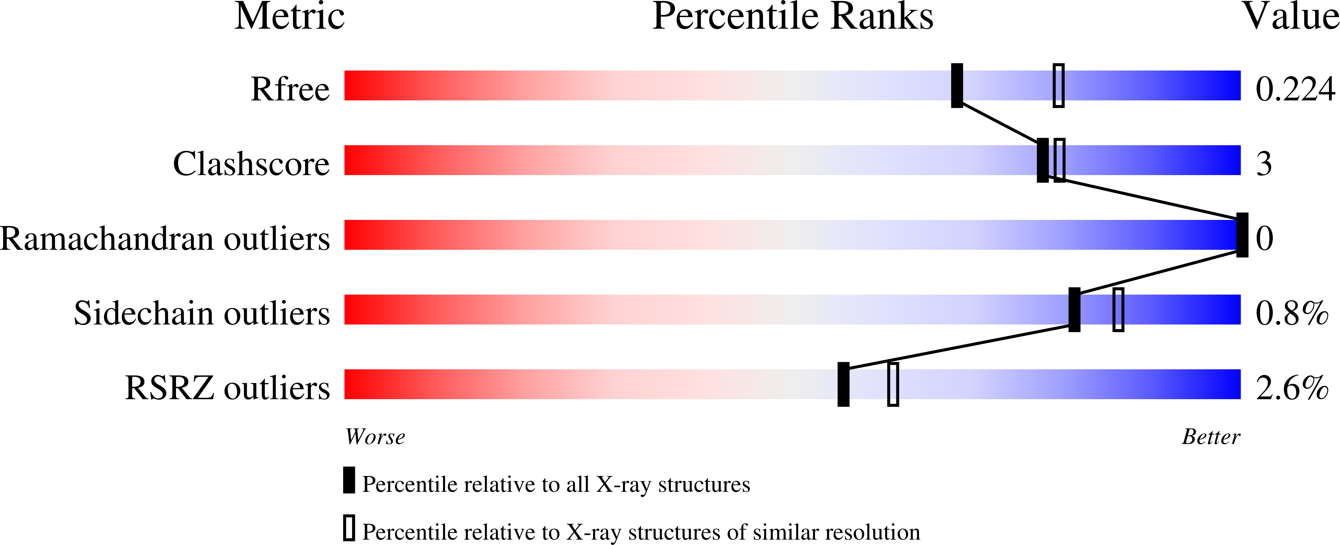
Experimental Data Snapshot
wwPDB Validation 3D Report Full Report
Entity ID: 1 | |||||
|---|---|---|---|---|---|
| Molecule | Chains | Sequence Length | Organism | Details | Image |
| Enoyl-CoA hydratase, EchA3 | 235 | Mycobacterium ulcerans Agy99 | Mutation(s): 0 Gene Names: echA3, MUL_0713 | 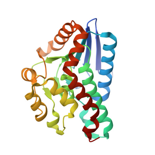 | |
UniProt | |||||
Find proteins for A0PLZ7 (Mycobacterium ulcerans (strain Agy99)) Explore A0PLZ7 Go to UniProtKB: A0PLZ7 | |||||
Entity Groups | |||||
| Sequence Clusters | 30% Identity50% Identity70% Identity90% Identity95% Identity100% Identity | ||||
| UniProt Group | A0PLZ7 | ||||
Sequence AnnotationsExpand | |||||
| |||||
| Length ( Å ) | Angle ( ˚ ) |
|---|---|
| a = 130.65 | α = 90 |
| b = 75.52 | β = 115.43 |
| c = 82.7 | γ = 90 |
| Software Name | Purpose |
|---|---|
| XDS | data reduction |
| XSCALE | data scaling |
| PHENIX | refinement |
| PDB_EXTRACT | data extraction |
| MoRDa | phasing |
| PHASER | phasing |
| Coot | model building |
RCSB PDB (citation) is hosted by
RCSB PDB is a member of the

