


Crystal Structure of Tyrosine-tRNA Synthetase from Acinetobacter baumannii with bound L-Tyrosine
Dranow, D.M., Horanyi, P.S., Lorimer, D.D., Edwards, T.E.To be published.
Experimental Data Snapshot
Entity ID: 1 | |||||
|---|---|---|---|---|---|
| Molecule | Chains | Sequence Length | Organism | Details | Image |
| Tyrosine--tRNA ligase | 408 | Acinetobacter baumannii | Mutation(s): 0 Gene Names: tyrS, A7A59_01455, A7A65_06465, A7M79_16285, A7M90_19385, A7N09_15205, APD06_07635, APD31_16655, B9X91_04915, B9X95_11600... EC: 6.1.1.1 | 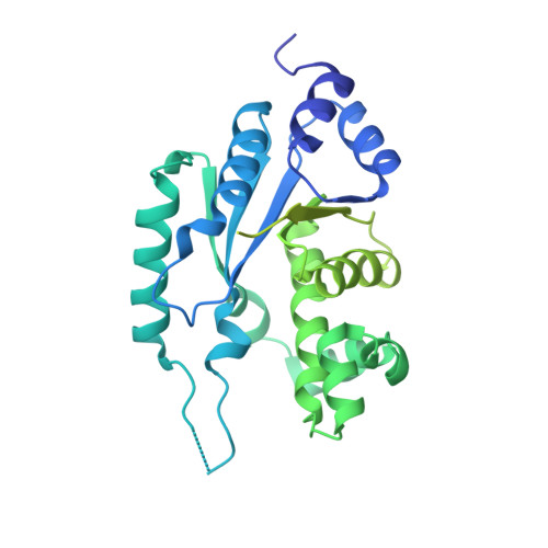 | |
UniProt | |||||
Find proteins for B0VML7 (Acinetobacter baumannii (strain SDF)) Explore B0VML7 Go to UniProtKB: B0VML7 | |||||
Entity Groups | |||||
| Sequence Clusters | 30% Identity50% Identity70% Identity90% Identity95% Identity100% Identity | ||||
| UniProt Group | B0VML7 | ||||
Sequence AnnotationsExpand | |||||
| |||||
| Ligands 3 Unique | |||||
|---|---|---|---|---|---|
| ID | Chains | Name / Formula / InChI Key | 2D Diagram | 3D Interactions | |
| TYR Query on TYR | C [auth A], E [auth B] | TYROSINE C9 H11 N O3 OUYCCCASQSFEME-QMMMGPOBSA-N |  | ||
| ACT Query on ACT | F [auth B] | ACETATE ION C2 H3 O2 QTBSBXVTEAMEQO-UHFFFAOYSA-M |  | ||
| MG Query on MG | D [auth A], G [auth B] | MAGNESIUM ION Mg JLVVSXFLKOJNIY-UHFFFAOYSA-N |  | ||
| Length ( Å ) | Angle ( ˚ ) |
|---|---|
| a = 48.26 | α = 87.65 |
| b = 49.8 | β = 107.11 |
| c = 73.37 | γ = 113.86 |
| Software Name | Purpose |
|---|---|
| XDS | data reduction |
| XSCALE | data scaling |
| PHASER | phasing |
| PHENIX | refinement |
| PDB_EXTRACT | data extraction |
RCSB PDB (citation) is hosted by
RCSB PDB is a member of the

