6B5K

Mycobacterium tuberculosis RmlA in complex with Mg/dTTP


Experimental Data Snapshot

  • Method: X-RAY DIFFRACTION
  • Resolution: 1.60 Å
  • R-Value Free: 0.227 
  • R-Value Work: 0.195 
  • R-Value Observed: 0.196 

wwPDB Validation   3D Report Full Report


Ligand Structure Quality Assessment 


This is version 1.2 of the entry. See complete history


Literature

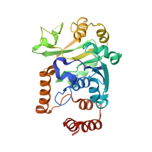
The structure of glucose-1-phosphate thymidylyltransferase from Mycobacterium tuberculosis reveals the location of an essential magnesium ion in the RmlA-type enzymes.

Brown, H.A.Thoden, J.B.Tipton, P.A.Holden, H.M.

(2018) Protein Sci 27: 441-450

  • DOI: https://doi.org/10.1002/pro.3333
  • Primary Citation of Related Structures:  
    6B5E, 6B5K

  • PubMed Abstract: 

    Tuberculosis, caused by the bacterium Mycobacterium tuberculosis, continues to be a major threat to populations worldwide. Whereas the disease is treatable, the drug regimen is arduous at best with the use of four antimicrobials over a six-month period. There is clearly a pressing need for the development of new therapeutics. One potential target for structure-based drug design is the enzyme RmlA, a glucose-1-phosphate thymidylyltransferase. This enzyme catalyzes the first step in the biosynthesis of l-rhamnose, which is a deoxysugar critical for the integrity of the bacterium's cell wall. Here, we report the X-ray structures of M. tuberculosis RmlA in complex with either dTTP or dTDP-glucose to 1.6 Å and 1.85 Å resolution, respectively. In the RmlA/dTTP complex, two magnesium ions were observed binding to the nucleotide, both ligated in octahedral coordination spheres. In the RmlA/dTDP-glucose complex, only a single magnesium ion was observed. Importantly, for RmlA-type enzymes with known three-dimensional structures, not one model shows the position of the magnesium ion bound to the nucleotide-linked sugar. As such, this investigation represents the first direct observation of the manner in which a magnesium ion is coordinated to the RmlA product and thus has important ramifications for structure-based drug design. In the past, molecular modeling procedures have been employed to derive a three-dimensional model of the M. tuberculosis RmlA for drug design. The X-ray structures presented herein provide a superior molecular scaffold for such endeavors in the treatment of one of the world's deadliest diseases.


  • Organizational Affiliation

    Department of Biochemistry, University of Wisconsin, Madison, WI, 53706, USA.


Macromolecules
Find similar proteins by:  (by identity cutoff)  |  3D Structure
Entity ID: 1
MoleculeChains Sequence LengthOrganismDetailsImage
Glucose-1-phosphate thymidylyltransferase
A, B
296Mycobacterium tuberculosis H37RvMutation(s): 0 
Gene Names: rmlArfbARv0334
EC: 2.7.7.24
UniProt
Find proteins for P9WH13 (Mycobacterium tuberculosis (strain ATCC 25618 / H37Rv))
Explore P9WH13 
Go to UniProtKB:  P9WH13
Entity Groups  
Sequence Clusters30% Identity50% Identity70% Identity90% Identity95% Identity100% Identity
UniProt GroupP9WH13
Sequence Annotations
Expand
  • Reference Sequence
Small Molecules
Ligands 4 Unique
IDChains Name / Formula / InChI Key2D Diagram3D Interactions
TTP
Query on TTP

Download Ideal Coordinates CCD File 
C [auth A],
J [auth B]
THYMIDINE-5'-TRIPHOSPHATE
C10 H17 N2 O14 P3
NHVNXKFIZYSCEB-XLPZGREQSA-N
TYD
Query on TYD

Download Ideal Coordinates CCD File 
F [auth A],
K [auth B]
THYMIDINE-5'-DIPHOSPHATE
C10 H16 N2 O11 P2
UJLXYODCHAELLY-XLPZGREQSA-N
EDO
Query on EDO

Download Ideal Coordinates CCD File 
G [auth A]1,2-ETHANEDIOL
C2 H6 O2
LYCAIKOWRPUZTN-UHFFFAOYSA-N
MG
Query on MG

Download Ideal Coordinates CCD File 
D [auth A],
E [auth A],
H [auth B],
I [auth B]
MAGNESIUM ION
Mg
JLVVSXFLKOJNIY-UHFFFAOYSA-N
Experimental Data & Validation

Experimental Data

  • Method: X-RAY DIFFRACTION
  • Resolution: 1.60 Å
  • R-Value Free: 0.227 
  • R-Value Work: 0.195 
  • R-Value Observed: 0.196 
  • Space Group: I 4
Unit Cell:
Length ( Å )Angle ( ˚ )
a = 96.403α = 90
b = 96.403β = 90
c = 151.924γ = 90
Software Package:
Software NamePurpose
REFMACrefinement
HKL-3000data reduction
HKL-3000data scaling
PHASERphasing

Structure Validation

View Full Validation Report


Ligand Structure Quality Assessment 


Entry History & Funding Information

Deposition Data


Funding OrganizationLocationGrant Number
National Institutes of Health/National Institute of General Medical Sciences (NIH/NIGMS)United StatesGM115921

Revision History  (Full details and data files)

  • Version 1.0: 2018-02-21
    Type: Initial release
  • Version 1.1: 2020-01-01
    Changes: Author supporting evidence
  • Version 1.2: 2023-10-04
    Changes: Data collection, Database references, Derived calculations, Refinement description