


Crystal structure of a 2,3,4,5-tetrahydropyridine-2,6-dicarboxylate N-succinyltransferase from Acinetobacter baumannii
Edwards, T.E., Horanyi, P.S., Lorimer, D.D., Seattle Structural Genomics Center for Infectious DiseaseTo be published.
Experimental Data Snapshot
wwPDB Validation 3D Report Full Report

Entity ID: 1 | |||||
|---|---|---|---|---|---|
| Molecule | Chains | Sequence Length | Organism | Details | Image |
| 2,3,4,5-tetrahydropyridine-2,6-dicarboxylate N-succinyltransferase | 281 | Acinetobacter baumannii | Mutation(s): 0 Gene Names: dapD EC: 2.3.1.117 | 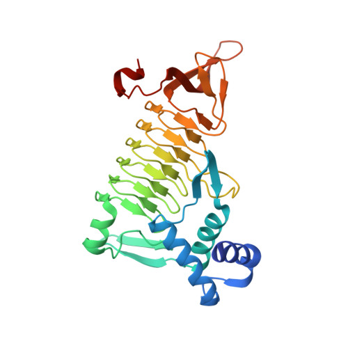 | |
UniProt | |||||
Find proteins for Q5DL43 (Acinetobacter baumannii) Explore Q5DL43 Go to UniProtKB: Q5DL43 | |||||
Entity Groups | |||||
| Sequence Clusters | 30% Identity50% Identity70% Identity90% Identity95% Identity100% Identity | ||||
| UniProt Group | Q5DL43 | ||||
Sequence AnnotationsExpand | |||||
| |||||
| Length ( Å ) | Angle ( ˚ ) |
|---|---|
| a = 64.36 | α = 90 |
| b = 83.21 | β = 90 |
| c = 148.04 | γ = 90 |
| Software Name | Purpose |
|---|---|
| PHENIX | refinement |
| XSCALE | data scaling |
| MOLREP | phasing |
| PDB_EXTRACT | data extraction |
| XDS | data reduction |
RCSB PDB (citation) is hosted by
RCSB PDB is a member of the

