6A2B

Crystal Structure of Xenopus laevis MHC I complex


Experimental Data Snapshot

  • Method: X-RAY DIFFRACTION
  • Resolution: 2.80 Å
  • R-Value Free: 0.264 
  • R-Value Work: 0.200 
  • R-Value Observed: 0.226 

wwPDB Validation   3D Report Full Report


This is version 1.3 of the entry. See complete history


Literature

A Glimpse of the Peptide Profile Presentation byXenopus laevisMHC Class I: Crystal Structure of pXela-UAA Reveals a Distinct Peptide-Binding Groove.

Ma, L.Zhang, N.Qu, Z.Liang, R.Zhang, L.Zhang, B.Meng, G.Dijkstra, J.M.Li, S.Xia, M.C.

(2020) J Immunol 204: 147-158

  • DOI: https://doi.org/10.4049/jimmunol.1900865
  • Primary Citation of Related Structures:  
    6A2B

  • PubMed Abstract: 

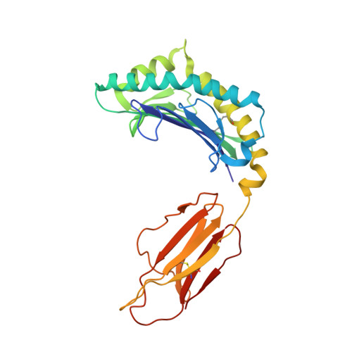
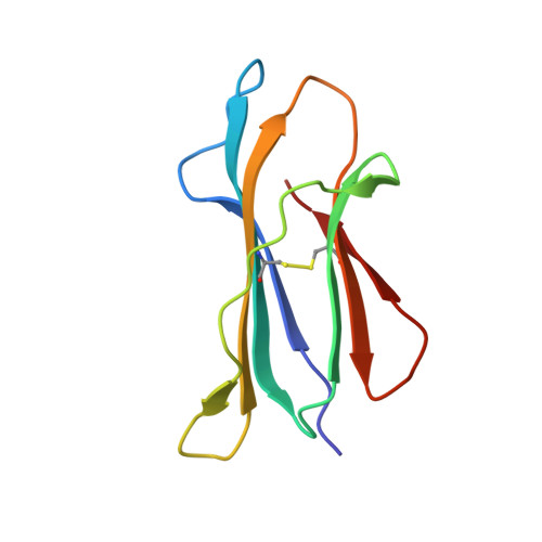
    The African clawed frog, Xenopus laevis , is a model species for amphibians. Before metamorphosis, tadpoles do not efficiently express the single classical MHC class I (MHC-I) molecule Xela -UAA, but after metamorphosis, adults express this molecule in abundance. To elucidate the Ag-presenting mechanism of Xela -UAA, in this study, the Xela -UAA structure complex (p Xela -UAAg) bound with a peptide from a synthetic random peptide library was determined. The amino acid homology between the Xela -UAA and MHC-I sequences of different species is <45%, and these differences are fully reflected in the three-dimensional structure of p Xela -UAAg. Because of polymorphisms and interspecific differences in amino acid sequences, p Xela -UAAg forms a distinct peptide-binding groove and presents a unique peptide profile. The most important feature of p Xela -UAAg is the two-amino acid insertion in the α2-helical region, which forms a protrusion of ∼3.8 Å that is involved in TCR docking. Comparison of peptide-MHC-I complex (pMHC-I) structures showed that only four amino acids in β2-microglobulin that were bound to MHC-I are conserved in almost all jawed vertebrates, and the most unique feature in nonmammalian pMHC-I molecules is that the AB loop bound β2-microglobulin. Additionally, the binding distance between pMHC-I and CD8 molecules in nonmammals is different from that in mammals. These unique features of p Xela -UAAg provide enhanced knowledge of T cell immunity and bridge the knowledge gap regarding the coevolutionary progression of the MHC-I complex from aquatic to terrestrial species.


  • Organizational Affiliation

    Department of Microbiology and Immunology, College of Veterinary Medicine, China Agricultural University, Beijing 100193, China.


Macromolecules
Find similar proteins by:  (by identity cutoff)  |  3D Structure
Entity ID: 1
MoleculeChains Sequence LengthOrganismDetailsImage
MHC class I antigen272Xenopus laevisMutation(s): 0 
UniProt
Find proteins for Q9TPA7 (Xenopus laevis)
Explore Q9TPA7 
Go to UniProtKB:  Q9TPA7
Entity Groups  
Sequence Clusters30% Identity50% Identity70% Identity90% Identity95% Identity100% Identity
UniProt GroupQ9TPA7
Sequence Annotations
Expand
  • Reference Sequence
Find similar proteins by:  (by identity cutoff)  |  3D Structure
Entity ID: 2
MoleculeChains Sequence LengthOrganismDetailsImage
Beta-2-microglobulin94Xenopus laevisMutation(s): 0 
Gene Names: b2m
UniProt
Find proteins for Q9IA97 (Xenopus laevis)
Explore Q9IA97 
Go to UniProtKB:  Q9IA97
Entity Groups  
Sequence Clusters30% Identity50% Identity70% Identity90% Identity95% Identity100% Identity
UniProt GroupQ9IA97
Sequence Annotations
Expand
  • Reference Sequence

Find similar proteins by:  Sequence   |   3D Structure  

Entity ID: 3
MoleculeChains Sequence LengthOrganismDetailsImage
TYR-MET-MET-PRO-ARG-HIS-TRP-PRO-ILE9Xenopus laevisMutation(s): 0 
Sequence Annotations
Expand
  • Reference Sequence
Experimental Data & Validation

Experimental Data

  • Method: X-RAY DIFFRACTION
  • Resolution: 2.80 Å
  • R-Value Free: 0.264 
  • R-Value Work: 0.200 
  • R-Value Observed: 0.226 
  • Space Group: P 21 21 21
Unit Cell:
Length ( Å )Angle ( ˚ )
a = 59.139α = 90
b = 68.22β = 90
c = 107.809γ = 90
Software Package:
Software NamePurpose
REFMACrefinement
HKL-2000data reduction
HKL-2000data scaling
PHASERphasing

Structure Validation

View Full Validation Report


Entry History 

Deposition Data

Revision History  (Full details and data files)

  • Version 1.0: 2019-10-23
    Type: Initial release
  • Version 1.1: 2019-12-11
    Changes: Database references
  • Version 1.2: 2020-01-01
    Changes: Database references
  • Version 1.3: 2023-11-22
    Changes: Data collection, Database references, Refinement description