


Structure of dipeptidyl peptidase III from Corallococcus sp. strain EGB
Zhang, H., Duan, Y.J., Li, Z.K., Liu, W.D., Huang, Y., Cui, Z.L.To be published.
Experimental Data Snapshot
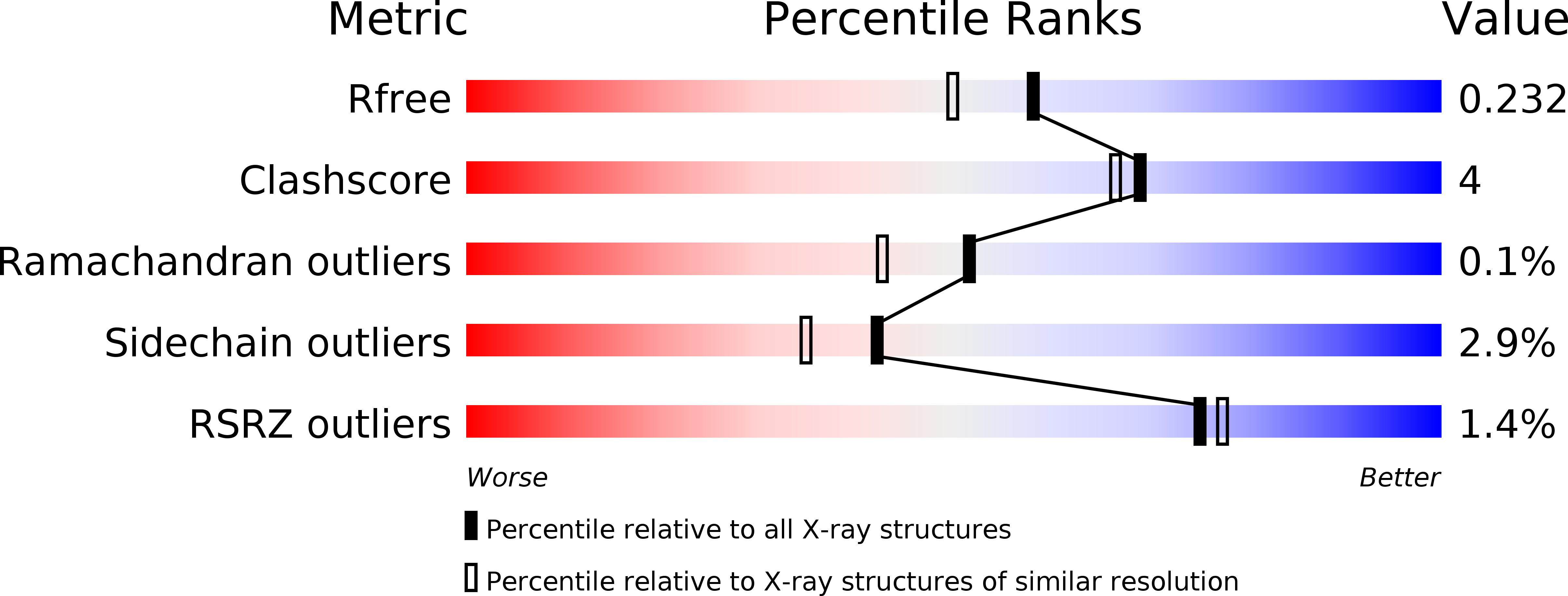
wwPDB Validation 3D Report Full Report
Entity ID: 1 | |||||
|---|---|---|---|---|---|
| Molecule | Chains | Sequence Length | Organism | Details | Image |
| dipeptidyl-peptidase III | 537 | Corallococcus sp. EGB | Mutation(s): 0 | 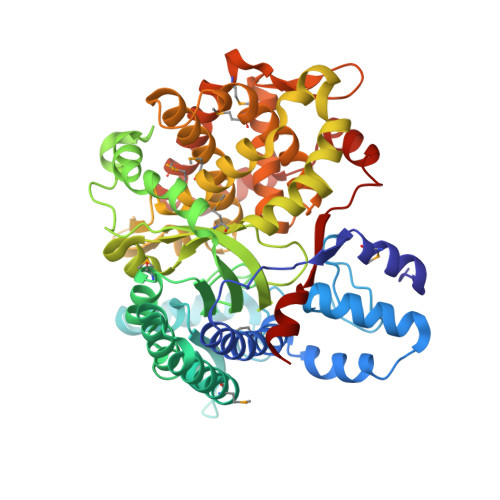 | |
UniProt | |||||
Find proteins for A0A5H1ZR28 (Corallococcus sp. EGB) Explore A0A5H1ZR28 Go to UniProtKB: A0A5H1ZR28 | |||||
Entity Groups | |||||
| Sequence Clusters | 30% Identity50% Identity70% Identity90% Identity95% Identity100% Identity | ||||
| UniProt Group | A0A5H1ZR28 | ||||
Sequence AnnotationsExpand | |||||
| |||||
| Ligands 1 Unique | |||||
|---|---|---|---|---|---|
| ID | Chains | Name / Formula / InChI Key | 2D Diagram | 3D Interactions | |
| ZN Query on ZN | C [auth A], D [auth B] | ZINC ION Zn PTFCDOFLOPIGGS-UHFFFAOYSA-N |  | ||
| Modified Residues 1 Unique | |||||
|---|---|---|---|---|---|
| ID | Chains | Type | Formula | 2D Diagram | Parent |
| MSE Query on MSE | A, B | L-PEPTIDE LINKING | C5 H11 N O2 Se |  | MET |
| Length ( Å ) | Angle ( ˚ ) |
|---|---|
| a = 58.141 | α = 90 |
| b = 77.85 | β = 90 |
| c = 228.294 | γ = 90 |
| Software Name | Purpose |
|---|---|
| REFMAC | refinement |
| MOSFLM | data reduction |
| XSCALE | data scaling |
| PHENIX | phasing |
RCSB PDB (citation) is hosted by
RCSB PDB is a member of the

