5ZUG

Structure of the bacterial acetate channel SatP


Experimental Data Snapshot

  • Method: X-RAY DIFFRACTION
  • Resolution: 2.80 Å
  • R-Value Free: 0.267 
  • R-Value Work: 0.225 
  • R-Value Observed: 0.227 

wwPDB Validation   3D Report Full Report


Ligand Structure Quality Assessment 


This is version 1.3 of the entry. See complete history


Literature

Crystal structure of the bacterial acetate transporter SatP reveals that it forms a hexameric channel.

Sun, P.Li, J.Zhang, X.Guan, Z.Xiao, Q.Zhao, C.Song, M.Zhou, Y.Mou, L.Ke, M.Guo, L.Geng, J.Deng, D.

(2018) J Biol Chem 293: 19492-19500

  • DOI: https://doi.org/10.1074/jbc.RA118.003876
  • Primary Citation of Related Structures:  
    5ZUG

  • PubMed Abstract: 

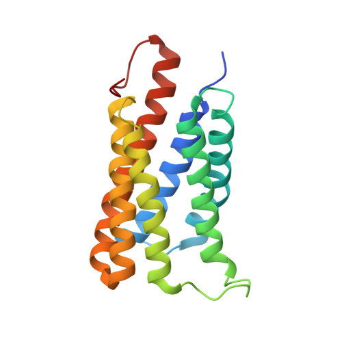
    Acetate is found ubiquitously in the natural environment and can be used as an exogenous carbon source by bacteria, fungi, and mammalian cells. A representative member of the acetate uptake transporter (AceTr) family named SatP (also yaaH) has been preliminarily identified as a succinate-acetate/proton symporter in Escherichia coli However, the molecular mechanism of acetate uptake by SatP still remains elusive. Here, we report the crystal structure of SatP from E. coli at 2.8 Å resolution, determined with a molecular replacement approach using a previously developed predicted model algorithm, which revealed a hexameric UreI-like channel structure. Structural analysis identified six transmembrane (TM) helices surrounding the central channel pore in each protomer and three conserved hydrophobic residues, FLY, located in the middle of the TM region for pore constriction. According to single-channel conductance recordings, performed with purified SatP reconstituted into lipid bilayer, three conserved polar residues in the TM1 facing to the periplasmic side are closely associated with acetate translocation activity. These analyses provide critical insights into the mechanism of acetate translocation in bacteria and a first glimpse of a structure of an AceTr family transporter.


  • Organizational Affiliation

    the School of Life Sciences, Tsinghua University, Beijing 100084, China, and.


Macromolecules
Find similar proteins by:  (by identity cutoff)  |  3D Structure
Entity ID: 1
MoleculeChains Sequence LengthOrganismDetailsImage
Succinate-acetate/proton symporter SatP
A, B, C, D, E
A, B, C, D, E, F
190Escherichia coli K-12Mutation(s): 0 
Gene Names: satPyaaHb0010JW0009
Membrane Entity: Yes 
UniProt
Find proteins for P0AC98 (Escherichia coli (strain K12))
Explore P0AC98 
Go to UniProtKB:  P0AC98
Entity Groups  
Sequence Clusters30% Identity50% Identity70% Identity90% Identity95% Identity100% Identity
UniProt GroupP0AC98
Sequence Annotations
Expand
  • Reference Sequence
Experimental Data & Validation

Experimental Data

  • Method: X-RAY DIFFRACTION
  • Resolution: 2.80 Å
  • R-Value Free: 0.267 
  • R-Value Work: 0.225 
  • R-Value Observed: 0.227 
  • Space Group: P 32
Unit Cell:
Length ( Å )Angle ( ˚ )
a = 99.504α = 90
b = 99.504β = 90
c = 137.824γ = 120
Software Package:
Software NamePurpose
PHENIXrefinement
HKL-3000data scaling
Cootmodel building
HKL-3000data reduction
PHENIXphasing

Structure Validation

View Full Validation Report


Ligand Structure Quality Assessment 


Entry History & Funding Information

Deposition Data


Funding OrganizationLocationGrant Number
National Institutes of Health/National Human Genome Research Institute (NIH/NHGRI)ChinaYFA052700
National Science Foundation (China)ChinaJQ007

Revision History  (Full details and data files)

  • Version 1.0: 2018-11-21
    Type: Initial release
  • Version 1.1: 2018-12-26
    Changes: Data collection, Database references
  • Version 1.2: 2020-07-29
    Type: Remediation
    Reason: Carbohydrate remediation
    Changes: Data collection, Derived calculations, Structure summary
  • Version 1.3: 2022-03-23
    Changes: Author supporting evidence, Database references, Structure summary