5ZNL

Crystal structure of PDE10A catalytic domain complexed with LHB-6


Experimental Data Snapshot

  • Method: X-RAY DIFFRACTION
  • Resolution: 2.80 Å
  • R-Value Free: 0.307 
  • R-Value Work: 0.234 
  • R-Value Observed: 0.238 

wwPDB Validation   3D Report Full Report


Ligand Structure Quality Assessment 


This is version 1.2 of the entry. See complete history


Literature

Absolute Binding Free Energy Calculation and Design of a Subnanomolar Inhibitor of Phosphodiesterase-10.

Li, Z.Huang, Y.Wu, Y.Chen, J.Wu, D.Zhan, C.G.Luo, H.B.

(2019) J Med Chem 62: 2099-2111

  • DOI: https://doi.org/10.1021/acs.jmedchem.8b01763
  • Primary Citation of Related Structures:  
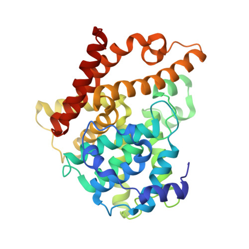
    5ZNL

  • PubMed Abstract: 

    Accurate prediction of absolute protein-ligand binding free energy could considerably enhance the success rate of structure-based drug design but is extremely challenging and time-consuming. Free energy perturbation (FEP) has been proven reliable but is limited to prediction of relative binding free energies of similar ligands (with only minor structural differences) in binding with a same drug target in practical drug design applications. Herein, a Gaussian algorithm-enhanced FEP (GA-FEP) protocol has been developed to enhance the FEP simulation performance, enabling to efficiently carry out the FEP simulations on vanishing the whole ligand and, thus, predict the absolute binding free energies (ABFEs). Using the GA-FEP protocol, the FEP simulations for the ABFE calculation (denoted as GA-FEP/ABFE) can achieve a satisfactory accuracy for both structurally similar and diverse ligands in a dataset of more than 100 receptor-ligand systems. Further, our GA-FEP/ABFE-guided lead optimization against phosphodiesterase-10 led to the discovery of a subnanomolar inhibitor (IC 50 = 0.87 nM, ∼2000-fold improvement in potency) with cocrystal confirmation.


  • Organizational Affiliation

    School of Pharmaceutical Sciences , Sun Yat-Sen University , Guangzhou 510006 , P.R. China.


Macromolecules
Find similar proteins by:  (by identity cutoff)  |  3D Structure
Entity ID: 1
MoleculeChains Sequence LengthOrganismDetailsImage
cAMP and cAMP-inhibited cGMP 3',5'-cyclic phosphodiesterase 10A
A, B
323Homo sapiensMutation(s): 0 
Gene Names: PDE10A
EC: 3.1.4.17 (PDB Primary Data), 3.1.4.35 (PDB Primary Data)
UniProt & NIH Common Fund Data Resources
Find proteins for Q9Y233 (Homo sapiens)
Explore Q9Y233 
Go to UniProtKB:  Q9Y233
PHAROS:  Q9Y233
GTEx:  ENSG00000112541 
Entity Groups  
Sequence Clusters30% Identity50% Identity70% Identity90% Identity95% Identity100% Identity
UniProt GroupQ9Y233
Sequence Annotations
Expand
  • Reference Sequence
Experimental Data & Validation

Experimental Data

  • Method: X-RAY DIFFRACTION
  • Resolution: 2.80 Å
  • R-Value Free: 0.307 
  • R-Value Work: 0.234 
  • R-Value Observed: 0.238 
  • Space Group: P 21 21 21
Unit Cell:
Length ( Å )Angle ( ˚ )
a = 49.151α = 90
b = 81.328β = 90
c = 158.207γ = 90
Software Package:
Software NamePurpose
REFMACrefinement
CrysalisProdata reduction
CrysalisProdata scaling
PHASERphasing

Structure Validation

View Full Validation Report


Ligand Structure Quality Assessment 


Entry History & Funding Information

Deposition Data


Funding OrganizationLocationGrant Number
National Natural Science Foundation of ChinaChina81522041, 81373258, 21572279, 21402243

Revision History  (Full details and data files)

  • Version 1.0: 2019-02-20
    Type: Initial release
  • Version 1.1: 2019-03-13
    Changes: Data collection, Database references
  • Version 1.2: 2023-11-22
    Changes: Data collection, Database references, Derived calculations, Refinement description