Crystal structures and kinetic analyses of N-acetylmannosamine-6-phosphate 2-epimerases from Fusobacterium nucleatum and Vibrio cholerae
Manjunath, L., Guntupalli, S.R., Currie, M.J., North, R.A., Dobson, R.C.J., Nayak, V., Subramanian, R.(2018) Acta Crystallogr F Struct Biol Commun 74: 431-440
- PubMed: 29969107
- DOI: https://doi.org/10.1107/S2053230X18008543
- Primary Citation of Related Structures:
5ZJB, 5ZJN, 5ZJP, 5ZKN - PubMed Abstract:
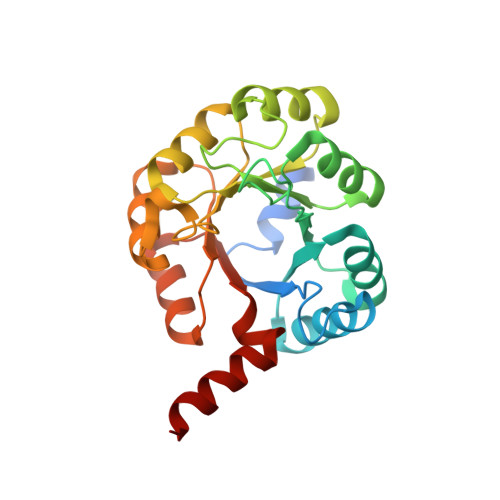
Sialic acids are nine-carbon sugars that are found abundantly on the cell surfaces of mammals as glycoprotein or glycolipid complexes. Several Gram-negative and Gram-positive bacteria have the ability to scavenge and catabolize sialic acids to use as a carbon source. This gives them an advantage in colonizing sialic acid-rich environments. The genes of the sialic acid catabolic pathway are generally present as the operon nanAKE. The third gene in the operon encodes the enzyme N-acetylmannosamine-6-phosphate 2-epimerase (NanE), which catalyzes the conversion of N-acetylmannosamine 6-phosphate to N-acetylglucosamine 6-phosphate, thus committing it to enter glycolysis. The NanE enzyme belongs to the isomerase class of enzymes possessing the triose phosphate isomerase (TIM) barrel fold. Here, comparative structural and functional characterizations of the NanE epimerases from two pathogenic Gram-negative bacteria, Fusobacterium nucleatum (Fn) and Vibrio cholerae (Vc), have been carried out. Structures of NanE from Vc (VcNanE) with and without ligand bound have been determined to 1.7 and 2.7 Å resolution, respectively. The structure of NanE from Fn (FnNanE) has been determined to 2.2 Å resolution. The enzymes show kinetic parameters that are consistent with those of Clostridium perfringens NanE. These studies allowed an evaluation of whether NanE may be a good drug target against these pathogenic bacteria.
Organizational Affiliation:
Institute for Stem Cell Biology and Regenerative Medicine, NCBS, GKVK Campus, Bellary Road, Bangalore, Karnataka 560 065, India.