


The Structure of cellobiose 2-epimerase from Spirochaeta thermophila DSM 6192
Feng, Y.H., Yang, R.J., Andrew, J.F.To be published.
Experimental Data Snapshot
Entity ID: 1 | |||||
|---|---|---|---|---|---|
| Molecule | Chains | Sequence Length | Organism | Details | Image |
| Cellobiose 2-epimerase | 404 | Spirochaeta thermophila DSM 6192 | Mutation(s): 0 Gene Names: STHERM_c00950 EC: 5.1.3.11 | 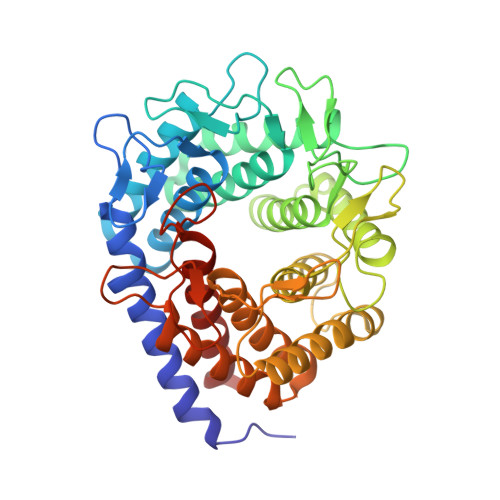 | |
UniProt | |||||
Find proteins for E0RU15 (Spirochaeta thermophila (strain ATCC 49972 / DSM 6192 / RI 19.B1)) Explore E0RU15 Go to UniProtKB: E0RU15 | |||||
Entity Groups | |||||
| Sequence Clusters | 30% Identity50% Identity70% Identity90% Identity95% Identity100% Identity | ||||
| UniProt Group | E0RU15 | ||||
Sequence AnnotationsExpand | |||||
| |||||
| Ligands 1 Unique | |||||
|---|---|---|---|---|---|
| ID | Chains | Name / Formula / InChI Key | 2D Diagram | 3D Interactions | |
| EDO (Subject of Investigation/LOI) Query on EDO | E [auth A], F [auth D] | 1,2-ETHANEDIOL C2 H6 O2 LYCAIKOWRPUZTN-UHFFFAOYSA-N |  | ||
| Length ( Å ) | Angle ( ˚ ) |
|---|---|
| a = 67.53 | α = 90 |
| b = 205.164 | β = 104.26 |
| c = 67.723 | γ = 90 |
| Software Name | Purpose |
|---|---|
| PHENIX | refinement |
| XDS | data reduction |
| XSCALE | data scaling |
| PHASER | phasing |
RCSB PDB (citation) is hosted by
RCSB PDB is a member of the

