5ZFS

Crystal structure of Arthrobacter globiformis M30 sugar epimerase which can produce D-allulose from D-fructose


Experimental Data Snapshot

  • Method: X-RAY DIFFRACTION
  • Resolution: 1.96 Å
  • R-Value Free: 0.211 
  • R-Value Work: 0.186 
  • R-Value Observed: 0.187 

wwPDB Validation   3D Report Full Report


This is version 1.1 of the entry. See complete history


Literature

X-ray structure of Arthrobacter globiformis M30 ketose 3-epimerase for the production of D-allulose from D-fructose.

Yoshida, H.Yoshihara, A.Gullapalli, P.K.Ohtani, K.Akimitsu, K.Izumori, K.Kamitori, S.

(2018) Acta Crystallogr F Struct Biol Commun 74: 669-676

  • DOI: https://doi.org/10.1107/S2053230X18011706
  • Primary Citation of Related Structures:  
    5ZFS

  • PubMed Abstract: 

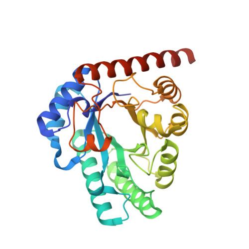
    The X-ray structure of ketose 3-epimerase from Arthrobacter globiformis M30, which was previously reported to be a D-allulose 3-epimerase (AgD-AE), was determined at 1.96 Å resolution. The crystal belonged to the hexagonal space group P6 5 22, with unit-cell parameters a = b = 103.98, c = 256.53 Å. The structure was solved by molecular replacement using the structure of Mesorhizobium loti L-ribulose 3-epimerase (MlL-RE), which has 41% sequence identity, as a search model. A hexagonal crystal contained two molecules in the asymmetric unit, and AgD-AE formed a homotetramer with twofold symmetry. The overall structure of AgD-AE was more similar to that of MlL-RE than to the known structures of D-psicose (alternative name D-allulose) 3-epimerases (D-PEs or D-AEs), although AgD-AE and MlL-RE have different substrate specificities. Both AgD-AE and MlL-RE have long helices in the C-terminal region that would contribute to the stability of the homotetramer. AgD-AE showed higher enzymatic activity for L-ribulose than D-allulose; however, AgD-AE is stable and is a unique useful enzyme for the production of D-allulose from D-fructose.


  • Organizational Affiliation

    Life Science Research Center and Faculty of Medicine, Kagawa University, 1750-1 Ikenobe, Miki-cho, Kita-gun, Kagawa 761-0793, Japan.


Macromolecules
Find similar proteins by:  (by identity cutoff)  |  3D Structure
Entity ID: 1
MoleculeChains Sequence LengthOrganismDetailsImage
D-allulose-3-epimerase
A, B
297Arthrobacter globiformisMutation(s): 0 
Gene Names: DAE
UniProt
Find proteins for A0A1L7NQ96 (Arthrobacter globiformis)
Explore A0A1L7NQ96 
Go to UniProtKB:  A0A1L7NQ96
Entity Groups  
Sequence Clusters30% Identity50% Identity70% Identity90% Identity95% Identity100% Identity
UniProt GroupA0A1L7NQ96
Sequence Annotations
Expand
  • Reference Sequence
Experimental Data & Validation

Experimental Data

  • Method: X-RAY DIFFRACTION
  • Resolution: 1.96 Å
  • R-Value Free: 0.211 
  • R-Value Work: 0.186 
  • R-Value Observed: 0.187 
  • Space Group: P 65 2 2
Unit Cell:
Length ( Å )Angle ( ˚ )
a = 103.98α = 90
b = 103.98β = 90
c = 256.53γ = 120
Software Package:
Software NamePurpose
REFMACrefinement
XDSdata reduction
XSCALEdata scaling
MOLREPphasing

Structure Validation

View Full Validation Report


Entry History & Funding Information

Deposition Data


Funding OrganizationLocationGrant Number
Japan Society for the Promotion of ScienceJapan16K07271

Revision History  (Full details and data files)

  • Version 1.0: 2018-10-24
    Type: Initial release
  • Version 1.1: 2023-11-22
    Changes: Data collection, Database references, Derived calculations, Refinement description