5ZCT

The crystal structure of the poly-alpha-L-glutamate peptides synthetase RimK at 2.05 angstrom resolution.


Experimental Data Snapshot

  • Method: X-RAY DIFFRACTION
  • Resolution: 2.05 Å
  • R-Value Free: 0.240 
  • R-Value Work: 0.196 
  • R-Value Observed: 0.198 

wwPDB Validation   3D Report Full Report


Ligand Structure Quality Assessment 


This is version 1.1 of the entry. See complete history


Literature

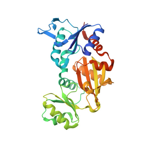
Structural polymorphism of the Escherichia coli poly-alpha-L-glutamate synthetase RimK

Arimura, Y.Kono, T.Kino, K.Kurumizaka, H.

(2018) Acta Crystallogr F Struct Biol Commun 74: 385-390

  • DOI: https://doi.org/10.1107/S2053230X18007689
  • Primary Citation of Related Structures:  
    5ZCT

  • PubMed Abstract: 

    Bacterial RimK is an enzyme that catalyzes the polyglutamylation of the C-terminus of ribosomal protein S6 and the synthesis of poly-α-L-glutamate peptides using L-glutamic acid. In the present study, the crystal structure of the Escherichia coli RimK protein complexed with the ATP analogue AMP-PNP was determined at 2.05 Å resolution. Two different conformations of RimK, closed and open forms, were observed in the crystals. The structural polymorphism revealed in this study provided important information to understand the mechanism by which RimK catalyzes the synthesis of poly-α-L-glutamate peptides and the polyglutamylation of ribosomal protein S6.


  • Organizational Affiliation

    Laboratory of Structural Biology, Graduate School of Advanced Science and Engineering, Waseda University, 2-2 Wakamatsu-cho, Shinjuku-ku, Tokyo 162-8480, Japan.


Macromolecules
Find similar proteins by:  (by identity cutoff)  |  3D Structure
Entity ID: 1
MoleculeChains Sequence LengthOrganismDetailsImage
Ribosomal protein S6--L-glutamate ligase
A, B, C, D, E
A, B, C, D, E, F, G, H
306Escherichia coli K-12Mutation(s): 0 
Gene Names: rimKb0852JW0836
EC: 6.3.2
UniProt
Find proteins for P0C0U4 (Escherichia coli (strain K12))
Explore P0C0U4 
Go to UniProtKB:  P0C0U4
Entity Groups  
Sequence Clusters30% Identity50% Identity70% Identity90% Identity95% Identity100% Identity
UniProt GroupP0C0U4
Sequence Annotations
Expand
  • Reference Sequence
Small Molecules
Ligands 3 Unique
IDChains Name / Formula / InChI Key2D Diagram3D Interactions
ANP
Query on ANP

Download Ideal Coordinates CCD File 
CA [auth F]
FA [auth G]
I [auth A]
JA [auth H]
N [auth B]
CA [auth F],
FA [auth G],
I [auth A],
JA [auth H],
N [auth B],
Q [auth C],
V [auth D],
Z [auth E]
PHOSPHOAMINOPHOSPHONIC ACID-ADENYLATE ESTER
C10 H17 N6 O12 P3
PVKSNHVPLWYQGJ-KQYNXXCUSA-N
SO4
Query on SO4

Download Ideal Coordinates CCD File 
BA [auth E]
EA [auth F]
HA [auth G]
IA [auth G]
K [auth A]
BA [auth E],
EA [auth F],
HA [auth G],
IA [auth G],
K [auth A],
L [auth A],
LA [auth H],
M [auth A],
P [auth B],
S [auth C],
T [auth C],
U [auth C],
X [auth D],
Y [auth E]
SULFATE ION
O4 S
QAOWNCQODCNURD-UHFFFAOYSA-L
MG
Query on MG

Download Ideal Coordinates CCD File 
AA [auth E]
DA [auth F]
GA [auth G]
J [auth A]
KA [auth H]
AA [auth E],
DA [auth F],
GA [auth G],
J [auth A],
KA [auth H],
O [auth B],
R [auth C],
W [auth D]
MAGNESIUM ION
Mg
JLVVSXFLKOJNIY-UHFFFAOYSA-N
Experimental Data & Validation

Experimental Data

  • Method: X-RAY DIFFRACTION
  • Resolution: 2.05 Å
  • R-Value Free: 0.240 
  • R-Value Work: 0.196 
  • R-Value Observed: 0.198 
  • Space Group: C 1 2 1
Unit Cell:
Length ( Å )Angle ( ˚ )
a = 212.618α = 90
b = 121.628β = 118.74
c = 119.741γ = 90
Software Package:
Software NamePurpose
HKL-2000data reduction
HKL-2000data scaling
PHASERphasing
PHENIXrefinement
PDB_EXTRACTdata extraction

Structure Validation

View Full Validation Report


Ligand Structure Quality Assessment 


Entry History 

Deposition Data

Revision History  (Full details and data files)

  • Version 1.0: 2018-07-25
    Type: Initial release
  • Version 1.1: 2023-11-22
    Changes: Data collection, Database references, Derived calculations, Refinement description