5ZCR

DSM5389 glycosyltrehalose synthase


Experimental Data Snapshot

  • Method: X-RAY DIFFRACTION
  • Resolution: 2.40 Å
  • R-Value Free: 0.248 
  • R-Value Work: 0.171 
  • R-Value Observed: 0.175 

wwPDB Validation   3D Report Full Report


This is version 1.1 of the entry. See complete history


Literature

Crystal structure of glycosyltrehalose synthase from Sulfolobus shibatae DSM5389

Okazaki, N.Blaber, M.Kuroki, R.Tamada, T.

(2018) Acta Crystallogr F Struct Biol Commun 74: 741-746

  • DOI: https://doi.org/10.1107/S2053230X1801453X
  • Primary Citation of Related Structures:  
    5ZCR

  • PubMed Abstract: 

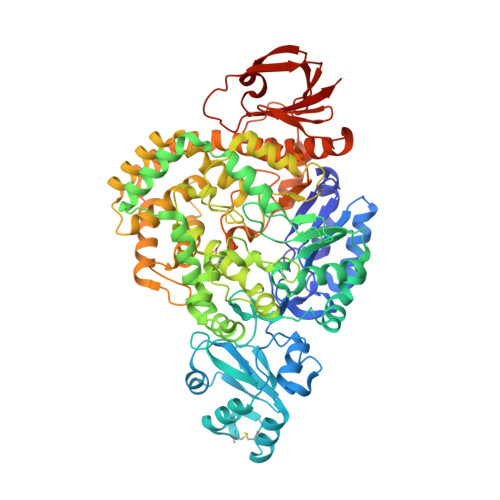
    Glycosyltrehalose synthase (GTSase) converts the glucosidic bond between the last two glucose residues of amylose from an α-1,4 bond to an α-1,1 bond, generating a nonreducing glycosyl trehaloside, in the first step of the biosynthesis of trehalose. To better understand the structural basis of the catalytic mechanism, the crystal structure of GTSase from the hyperthermophilic archaeon Sulfolobus shibatae DSM5389 (5389-GTSase) has been determined to 2.4 Å resolution by X-ray crystallography. The structure of 5389-GTSase can be divided into five domains. The central domain contains the (β/α) 8 -barrel fold that is conserved as the catalytic domain in the α-amylase family. Three invariant catalytic carboxylic amino acids in the α-amylase family are also found in GTSase at positions Asp241, Glu269 and Asp460 in the catalytic domain. The shape of the catalytic cavity and the pocket size at the bottom of the cavity correspond to the intramolecular transglycosylation mechanism proposed from previous enzymatic studies.


  • Organizational Affiliation

    Neutron Science and Technology Center, Comprehensive Research Organization for Science and Society, 162-1 Shirakata, Tokai, Ibaraki 319-1106, Japan.


Macromolecules
Find similar proteins by:  (by identity cutoff)  |  3D Structure
Entity ID: 1
MoleculeChains Sequence LengthOrganismDetailsImage
Maltooligosyl trehalose synthase
A, B
728Saccharolobus shibatae B12Mutation(s): 2 
Gene Names: treY
UniProt
Find proteins for Q7LYV2 (Saccharolobus shibatae (strain ATCC 51178 / DSM 5389 / JCM 8931 / NBRC 15437 / B12))
Explore Q7LYV2 
Go to UniProtKB:  Q7LYV2
Entity Groups  
Sequence Clusters30% Identity50% Identity70% Identity90% Identity95% Identity100% Identity
UniProt GroupQ7LYV2
Sequence Annotations
Expand
  • Reference Sequence
Small Molecules
Ligands 2 Unique
IDChains Name / Formula / InChI Key2D Diagram3D Interactions
GOL
Query on GOL

Download Ideal Coordinates CCD File 
D [auth A]
E [auth A]
F [auth A]
G [auth A]
H [auth A]
D [auth A],
E [auth A],
F [auth A],
G [auth A],
H [auth A],
I [auth A],
J [auth A],
L [auth B],
M [auth B],
N [auth B],
O [auth B]
GLYCEROL
C3 H8 O3
PEDCQBHIVMGVHV-UHFFFAOYSA-N
MG
Query on MG

Download Ideal Coordinates CCD File 
C [auth A],
K [auth B]
MAGNESIUM ION
Mg
JLVVSXFLKOJNIY-UHFFFAOYSA-N
Experimental Data & Validation

Experimental Data

  • Method: X-RAY DIFFRACTION
  • Resolution: 2.40 Å
  • R-Value Free: 0.248 
  • R-Value Work: 0.171 
  • R-Value Observed: 0.175 
  • Space Group: P 1 21 1
Unit Cell:
Length ( Å )Angle ( ˚ )
a = 71.132α = 90
b = 84.567β = 103.75
c = 128.662γ = 90
Software Package:
Software NamePurpose
PHENIXrefinement
HKL-2000data reduction
HKL-2000data scaling
MOLREPphasing

Structure Validation

View Full Validation Report


Entry History 

Deposition Data

Revision History  (Full details and data files)

  • Version 1.0: 2018-11-21
    Type: Initial release
  • Version 1.1: 2023-11-22
    Changes: Data collection, Database references, Derived calculations, Refinement description