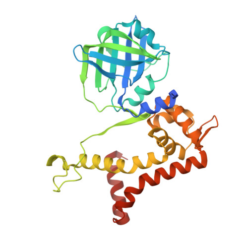
Crystal structure of the flavin reductase of Acinetobacter baumannii p-hydroxyphenylacetate 3-hydroxylase (HPAH) and identification of amino acid residues underlying its regulation by aromatic ligands
Yuenyao, A., Petchyam, N., Kamonsutthipaijit, N., Chaiyen, P., Pakotiprapha, D.(2018) Arch Biochem Biophys 653: 24-38
- PubMed: 29940152
- DOI: https://doi.org/10.1016/j.abb.2018.06.010
- Primary Citation of Related Structures:
5ZC2 - PubMed Abstract:
The first step in the degradation of p-hydroxyphenylacetic acid (HPA) is catalyzed by the two-component enzyme p-hydroxyphenylacetate 3-hydroxylase (HPAH). The two components of Acinetobacter baumannii HPAH are known as C 1 and C 2 , respectively. C 1 is a flavin reductase that uses NADH to generate reduced flavin mononucleotide (FMNH - ), which is used by C 2 in the hydroxylation of HPA. Interestingly, although HPA is not directly involved in the reaction catalyzed by C 1 , the presence of HPA dramatically increases the FMN reduction rate. Amino acid sequence analysis revealed that C 1 contains two domains: an N-terminal flavin reductase domain, and a C-terminal MarR domain. Although MarR proteins typically function as transcription regulators, the MarR domain of C 1 was found to play an auto-inhibitory role. Here, we report a crystal structure of C 1 and small-angle X-ray scattering (SAXS) studies that revealed that C 1 undergoes a substantial conformational change in the presence of HPA, concomitant with the increase in the rate of flavin reduction. Amino acid residues that are important for HPA binding and regulation of C 1 activity were identified by site-directed mutagenesis. Amino acid sequence similarity analysis revealed several as yet uncharacterized flavin reductases with N- or C-terminal fusions.
Organizational Affiliation:
Department of Biochemistry and Center for Excellence in Protein and Enzyme Technology Faculty of Science, Mahidol University, Bangkok, 10400, Thailand.