5Z79

Crystal Structure Analysis of the HPPK-DHPS in complex with substrates


Experimental Data Snapshot

  • Method: X-RAY DIFFRACTION
  • Resolution: 2.90 Å
  • R-Value Free: 0.285 
  • R-Value Work: 0.242 
  • R-Value Observed: 0.243 

wwPDB Validation   3D Report Full Report


Ligand Structure Quality Assessment 


This is version 1.4 of the entry. See complete history

Re-refinement Note

A newer entry is available that reflects an alternative modeling of the original data: 5Z79


Literature

Structure of 6-hydroxymethyl-7,8-dihydropterin pyrophosphokinase-dihydropteroate synthase fromPlasmodium vivaxsheds light on drug resistance

Yogavel, M.Nettleship, J.E.Sharma, A.Harlos, K.Jamwal, A.Chaturvedi, R.Sharma, M.Jain, V.Chhibber-Goel, J.Sharma, A.

(2018) J Biol Chem 293: 14962-14972

  • DOI: https://doi.org/10.1074/jbc.RA118.004558
  • Primary Citation of Related Structures:  
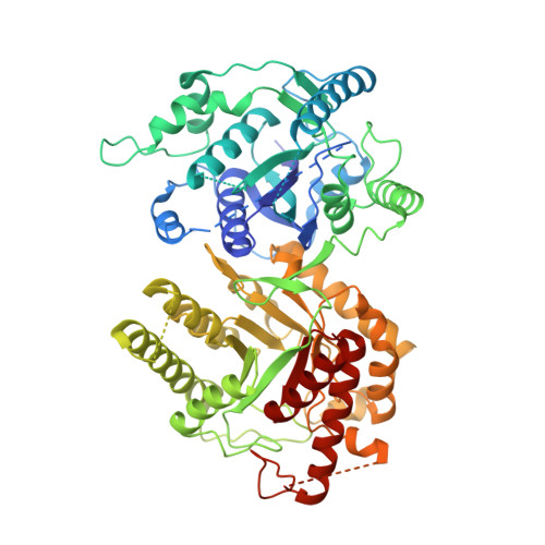
    5Z79

  • PubMed Abstract: 

    The genomes of the malaria-causing Plasmodium parasites encode a protein fused of 6-hydroxymethyl-7,8-dihydropterin pyrophosphokinase (HPPK) and dihydropteroate synthase (DHPS) domains that catalyze sequential reactions in the folate biosynthetic pathway. Whereas higher organisms derive folate from their diet and lack the enzymes for its synthesis, most eubacteria and a number of lower eukaryotes including malaria parasites synthesize tetrahydrofolate via DHPS. Plasmodium falciparum ( Pf ) and Plasmodium vivax ( Pv ) HPPK-DHPSs are currently targets of drugs like sulfadoxine (SDX). The SDX effectiveness as an antimalarial drug is increasingly diminished by the rise and spread of drug-resistant mutations. Here, we present the crystal structure of Pv HPPK-DHPS in complex with four substrates/analogs, revealing the bifunctional Pv HPPK-DHPS architecture in an unprecedented state of enzymatic activation. SDX's effect on HPPK-DHPS is due to 4-amino benzoic acid ( p ABA) mimicry, and the Pv HPPK-DHPS structure sheds light on the SDX-binding cavity, as well as on mutations that effect SDX potency. We mapped five dominant drug resistance mutations in Pv HPPK-DHPS: S382A, A383G, K512E/D, A553G, and V585A, most of which occur individually or in clusters proximal to the p ABA-binding site. We found that these resistance mutations subtly alter the intricate enzyme/ p ABA/SDX interactions such that DHPS affinity for p ABA is diminished only moderately, but its affinity for SDX is changed substantially. In conclusion, the Pv HPPK-DHPS structure rationalizes and unravels the structural bases for SDX resistance mutations and highlights architectural features in HPPK-DHPSs from malaria parasites that can form the basis for developing next-generation anti-folate agents to combat malaria parasites.


  • Organizational Affiliation

    From the Molecular Medicine-Structural Parasitology Group, International Centre for Genetic Engineering and Biotechnology, New Delhi 110067, India, myogavel@gmail.com.


Macromolecules
Find similar proteins by:  (by identity cutoff)  |  3D Structure
Entity ID: 1
MoleculeChains Sequence LengthOrganismDetailsImage
Hydroxymethyldihydropterin pyrophosphokinase-dihydropteroate synthase, putative
A, B, C, D, E
A, B, C, D, E, F
717Plasmodium vivaxMutation(s): 0 
EC: 2.5.1.15 (PDB Primary Data), 2.7.6.3 (PDB Primary Data)
UniProt
Find proteins for A5JZS1 (Plasmodium vivax (strain Salvador I))
Explore A5JZS1 
Go to UniProtKB:  A5JZS1
Entity Groups  
Sequence Clusters30% Identity50% Identity70% Identity90% Identity95% Identity100% Identity
UniProt GroupA5JZS1
Sequence Annotations
Expand
  • Reference Sequence
Small Molecules
Ligands 6 Unique
IDChains Name / Formula / InChI Key2D Diagram3D Interactions
APC
Query on APC

Download Ideal Coordinates CCD File 
H [auth A],
P [auth C]
DIPHOSPHOMETHYLPHOSPHONIC ACID ADENOSYL ESTER
C11 H18 N5 O12 P3
CAWZRIXWFRFUQB-IOSLPCCCSA-N
HH2
Query on HH2

Download Ideal Coordinates CCD File 
CA [auth F]
G [auth A]
L [auth B]
O [auth C]
T [auth D]
CA [auth F],
G [auth A],
L [auth B],
O [auth C],
T [auth D],
Y [auth E]
6-HYDROXYMETHYLPTERIN-DIPHOSPHATE
C7 H9 N5 O8 P2
AMDUVUKDRBIVAH-UHFFFAOYSA-N
5AD
Query on 5AD

Download Ideal Coordinates CCD File 
M [auth B],
U [auth D]
5'-DEOXYADENOSINE
C10 H13 N5 O3
XGYIMTFOTBMPFP-KQYNXXCUSA-N
PE0
Query on PE0

Download Ideal Coordinates CCD File 
BA [auth E],
K [auth A],
S [auth C],
X [auth D]
PTERINE
C6 H5 N5 O
HNXQXTQTPAJEJL-UHFFFAOYSA-N
PAB
Query on PAB

Download Ideal Coordinates CCD File 
DA [auth F]
I [auth A]
N [auth B]
Q [auth C]
V [auth D]
DA [auth F],
I [auth A],
N [auth B],
Q [auth C],
V [auth D],
Z [auth E]
4-AMINOBENZOIC ACID
C7 H7 N O2
ALYNCZNDIQEVRV-UHFFFAOYSA-N
MG
Query on MG

Download Ideal Coordinates CCD File 
AA [auth E],
J [auth A],
R [auth C],
W [auth D]
MAGNESIUM ION
Mg
JLVVSXFLKOJNIY-UHFFFAOYSA-N
Experimental Data & Validation

Experimental Data

  • Method: X-RAY DIFFRACTION
  • Resolution: 2.90 Å
  • R-Value Free: 0.285 
  • R-Value Work: 0.242 
  • R-Value Observed: 0.243 
  • Space Group: P 1 21 1
Unit Cell:
Length ( Å )Angle ( ˚ )
a = 136.049α = 90
b = 113.892β = 94.24
c = 172.387γ = 90
Software Package:
Software NamePurpose
REFMACrefinement
PHASERphasing
PDB_EXTRACTdata extraction
xia2data reduction
DIALSdata scaling

Structure Validation

View Full Validation Report


Ligand Structure Quality Assessment 


Entry History 

Deposition Data

Revision History  (Full details and data files)

  • Version 1.0: 2018-08-29
    Type: Initial release
  • Version 1.1: 2018-10-10
    Changes: Data collection, Database references, Structure summary
  • Version 1.2: 2019-12-18
    Changes: Data collection, Structure summary
  • Version 1.3: 2019-12-25
    Changes: Data collection
  • Version 1.4: 2023-11-22
    Changes: Data collection, Database references, Derived calculations, Refinement description