5Z5K

Structure of the DCC-Draxin complex

  • Classification: APOPTOSIS/INBITITOR
  • Organism(s): Rattus norvegicus
  • Expression System: Homo sapiens
  • Mutation(s): No 

  • Deposited: 2018-01-18 Released: 2018-06-20 
  • Deposition Author(s): Liu, Y., Xiao, J., Wang, J.
  • Funding Organization(s): National Science Foundation (China), National Institutes of Health/National Heart, Lung, and Blood Institute (NIH/NHLBI)

Experimental Data Snapshot

  • Method: X-RAY DIFFRACTION
  • Resolution: 2.49 Å
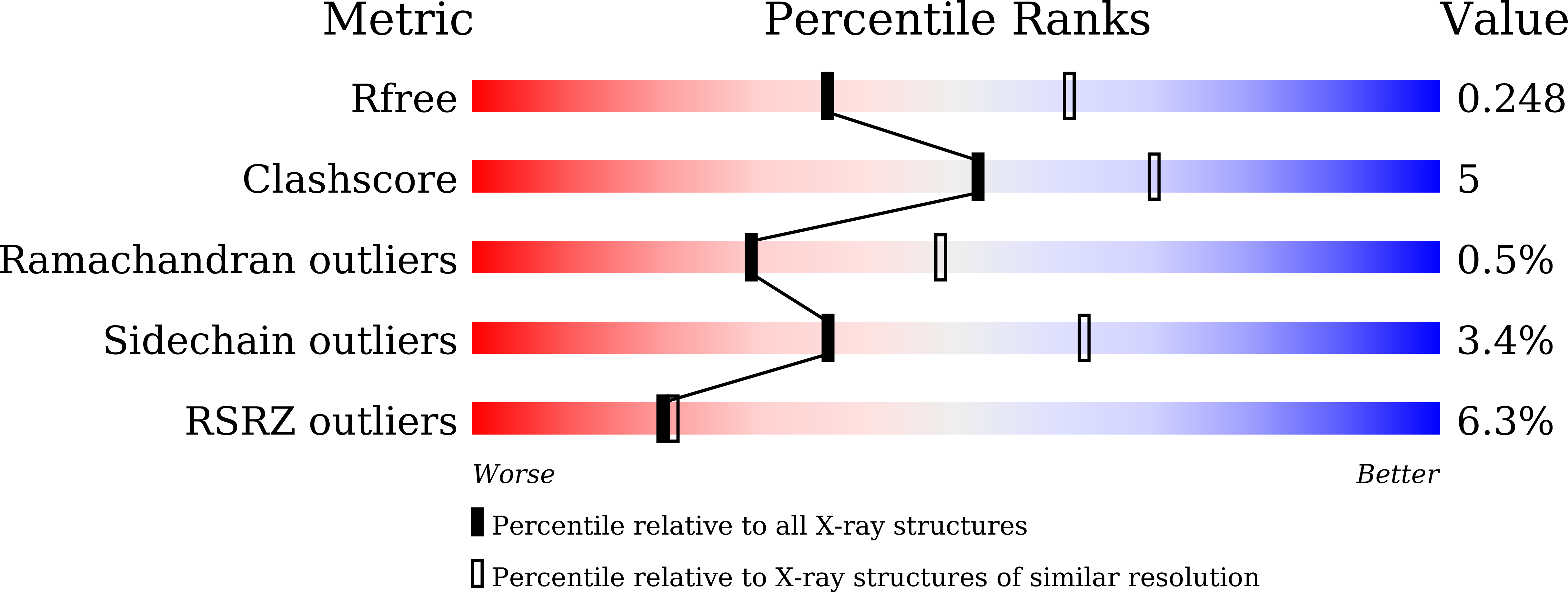
  • R-Value Free: 0.247 
  • R-Value Work: 0.212 
  • R-Value Observed: 0.214 

wwPDB Validation   3D Report Full Report


Ligand Structure Quality Assessment 


This is version 2.1 of the entry. See complete history


Literature

Structural Basis for Draxin-Modulated Axon Guidance and Fasciculation by Netrin-1 through DCC.

Liu, Y.Bhowmick, T.Liu, Y.Gao, X.Mertens, H.D.T.Svergun, D.I.Xiao, J.Zhang, Y.Wang, J.H.Meijers, R.

(2018) Neuron 97: 1261-1267.e4

  • DOI: https://doi.org/10.1016/j.neuron.2018.02.010
  • Primary Citation of Related Structures:  
    5Z5K, 6FKQ

  • PubMed Abstract: 

    Axon guidance involves the spatiotemporal interplay between guidance cues and membrane-bound cell-surface receptors, present on the growth cone of the axon. Netrin-1 is a prototypical guidance cue that binds to deleted in colorectal cancer (DCC), and it has been proposed that the guidance cue Draxin modulates this interaction. Here, we present structural snapshots of Draxin/DCC and Draxin/Netrin-1 complexes, revealing a triangular relationship that affects Netrin-mediated haptotaxis and fasciculation. Draxin interacts with DCC through the N-terminal four immunoglobulin domains, and Netrin-1 through the EGF-3 domain, in the same region where DCC binds. Netrin-1 and DCC bind to adjacent sites on Draxin, which appears to capture Netrin-1 and tether it to the DCC receptor. We propose the conformational flexibility of the single-pass membrane receptor DCC is used to promote fasciculation and regulate axon guidance through concerted Netrin-1/Draxin binding. VIDEO ABSTRACT.


  • Organizational Affiliation

    State Key Laboratory of Membrane Biology, College of Life Sciences, Peking University, Beijing 100871, China.


Macromolecules
Find similar proteins by:  (by identity cutoff)  |  3D Structure
Entity ID: 1
MoleculeChains Sequence LengthOrganismDetailsImage
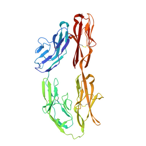
Netrin receptor DCC380Rattus norvegicusMutation(s): 0 
Gene Names: Dcc
UniProt
Find proteins for Q63155 (Rattus norvegicus)
Explore Q63155 
Go to UniProtKB:  Q63155
Entity Groups  
Sequence Clusters30% Identity50% Identity70% Identity90% Identity95% Identity100% Identity
UniProt GroupQ63155
Sequence Annotations
Expand
  • Reference Sequence
Find similar proteins by:  (by identity cutoff)  |  3D Structure
Entity ID: 2
MoleculeChains Sequence LengthOrganismDetailsImage
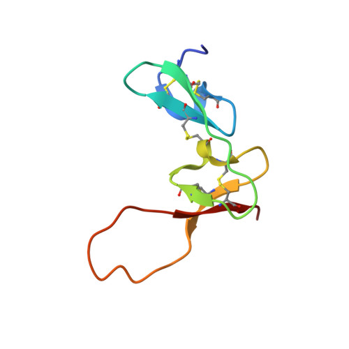
Draxin66Rattus norvegicusMutation(s): 0 
Gene Names: DraxinDRAXIN
UniProt
Find proteins for D3ZDG4 (Rattus norvegicus)
Explore D3ZDG4 
Go to UniProtKB:  D3ZDG4
Entity Groups  
Sequence Clusters30% Identity50% Identity70% Identity90% Identity95% Identity100% Identity
UniProt GroupD3ZDG4
Sequence Annotations
Expand
  • Reference Sequence
Oligosaccharides

Help

Entity ID: 3
MoleculeChains Length2D Diagram Glycosylation3D Interactions
2-acetamido-2-deoxy-beta-D-glucopyranose-(1-4)-2-acetamido-2-deoxy-beta-D-glucopyranose
C, D, E
2N-Glycosylation
Glycosylation Resources
GlyTouCan:  G42666HT
GlyCosmos:  G42666HT
GlyGen:  G42666HT
Small Molecules
Ligands 1 Unique
IDChains Name / Formula / InChI Key2D Diagram3D Interactions
NAG
Query on NAG

Download Ideal Coordinates CCD File 
F [auth A]2-acetamido-2-deoxy-beta-D-glucopyranose
C8 H15 N O6
OVRNDRQMDRJTHS-FMDGEEDCSA-N
Experimental Data & Validation

Experimental Data

  • Method: X-RAY DIFFRACTION
  • Resolution: 2.49 Å
  • R-Value Free: 0.247 
  • R-Value Work: 0.212 
  • R-Value Observed: 0.214 
  • Space Group: P 65
Unit Cell:
Length ( Å )Angle ( ˚ )
a = 108.069α = 90
b = 108.069β = 90
c = 130.266γ = 120
Software Package:
Software NamePurpose
SCALEPACKdata scaling
PHASERphasing
PHENIXrefinement
PDB_EXTRACTdata extraction

Structure Validation

View Full Validation Report


Ligand Structure Quality Assessment 


Entry History & Funding Information

Deposition Data


Funding OrganizationLocationGrant Number
National Science Foundation (China)China31570735
National Institutes of Health/National Heart, Lung, and Blood Institute (NIH/NHLBI)United StatesHL103526

Revision History  (Full details and data files)

  • Version 1.0: 2018-06-20
    Type: Initial release
  • Version 2.0: 2020-07-29
    Type: Remediation
    Reason: Carbohydrate remediation
    Changes: Advisory, Atomic model, Data collection, Derived calculations, Structure summary
  • Version 2.1: 2021-03-10
    Changes: Author supporting evidence, Structure summary