5Z51

Helicase binding domain of primase from Mycobacterium tuberculosis


Experimental Data Snapshot

  • Method: X-RAY DIFFRACTION
  • Resolution: 1.58 Å
  • R-Value Free: 0.209 
  • R-Value Work: 0.181 
  • R-Value Observed: 0.183 

wwPDB Validation   3D Report Full Report


This is version 1.0 of the entry. See complete history


Literature

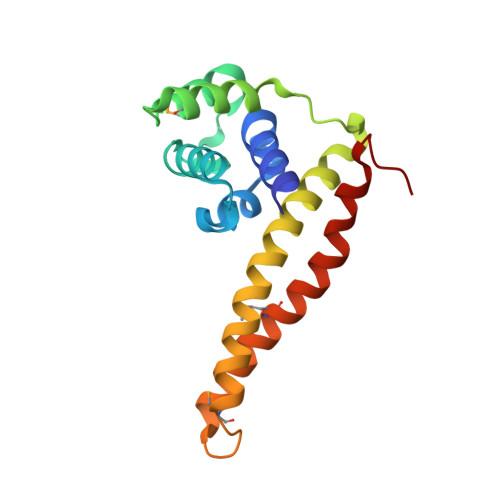
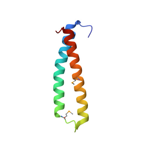
Structural insights into the interaction of helicase and primase inMycobacterium tuberculosis.

Sharma, D.P.Vijayan, R.Rehman, S.A.A.Gourinath, S.

(2018) Biochem J 475: 3493-3509

  • DOI: https://doi.org/10.1042/BCJ20180673
  • Primary Citation of Related Structures:  
    5Z51

  • PubMed Abstract: 

    The helicase-primase interaction is an essential event in DNA replication and is mediated by the highly variable C-terminal domain of primase (DnaG) and N-terminal domain of helicase (DnaB). To understand the functional conservation despite the low sequence homology of the DnaB-binding domains of DnaGs of eubacteria, we determined the crystal structure of the helicase-binding domain of DnaG from Mycobacterium tuberculosis ( Mt DnaG-CTD) and did so to a resolution of 1.58 Å. We observed the overall structure of Mt DnaG-CTD to consist of two subdomains, the N-terminal globular region (GR) and the C-terminal helical hairpin region (HHR), connected by a small loop. Despite differences in some of its helices, the globular region was found to have broadly similar arrangements across the species, whereas the helical hairpins showed different orientations. To gain insights into the crucial helicase-primase interaction in M. tuberculosis , a complex was modeled using the Mt DnaG-CTD and Mt DnaB-NTD crystal structures. Two nonconserved hydrophobic residues (Ile605 and Phe615) of Mt DnaG were identified as potential key residues interacting with Mt DnaB. Biosensor-binding studies showed a significant decrease in the binding affinity of Mt DnaB-NTD with the Ile605Ala mutant of Mt DnaG-CTD compared with native Mt DnaG-CTD. The loop, connecting the two helices of the HHR, was concluded to be largely responsible for the stability of the DnaB-DnaG complex. Also, Mt DnaB-NTD showed micromolar affinity with DnaG-CTDs from Escherichia coli and Helicobacter pylori and unstable binding with DnaG-CTD from Vibrio cholerae The interacting domains of both DnaG and DnaB demonstrate the species-specific evolution of the replication initiation system.


  • Organizational Affiliation

    School of Life Sciences, Jawaharlal Nehru University, Delhi 110067, India.


Macromolecules
Find similar proteins by:  (by identity cutoff)  |  3D Structure
Entity ID: 1
MoleculeChains Sequence LengthOrganismDetailsImage
DNA primase162Mycobacterium tuberculosis H37RvMutation(s): 0 
Gene Names: dnaGRv2343cMTCY98.12c
EC: 2.7.7
UniProt
Find proteins for P9WNW1 (Mycobacterium tuberculosis (strain ATCC 25618 / H37Rv))
Explore P9WNW1 
Go to UniProtKB:  P9WNW1
Entity Groups  
Sequence Clusters30% Identity50% Identity70% Identity90% Identity95% Identity100% Identity
UniProt GroupP9WNW1
Sequence Annotations
Expand
  • Reference Sequence
Find similar proteins by:  (by identity cutoff)  |  3D Structure
Entity ID: 2
MoleculeChains Sequence LengthOrganismDetailsImage
DNA primase71Mycobacterium tuberculosis H37RvMutation(s): 0 
Gene Names: dnaGRv2343cMTCY98.12c
EC: 2.7.7
UniProt
Find proteins for P9WNW1 (Mycobacterium tuberculosis (strain ATCC 25618 / H37Rv))
Explore P9WNW1 
Go to UniProtKB:  P9WNW1
Entity Groups  
Sequence Clusters30% Identity50% Identity70% Identity90% Identity95% Identity100% Identity
UniProt GroupP9WNW1
Sequence Annotations
Expand
  • Reference Sequence
Small Molecules
Ligands 2 Unique
IDChains Name / Formula / InChI Key2D Diagram3D Interactions
PEG
Query on PEG

Download Ideal Coordinates CCD File 
C [auth A]DI(HYDROXYETHYL)ETHER
C4 H10 O3
MTHSVFCYNBDYFN-UHFFFAOYSA-N
ACT
Query on ACT

Download Ideal Coordinates CCD File 
D [auth B]ACETATE ION
C2 H3 O2
QTBSBXVTEAMEQO-UHFFFAOYSA-M
Modified Residues  1 Unique
IDChains TypeFormula2D DiagramParent
MSE
Query on MSE
A
L-PEPTIDE LINKINGC5 H11 N O2 SeMET
Experimental Data & Validation

Experimental Data

  • Method: X-RAY DIFFRACTION
  • Resolution: 1.58 Å
  • R-Value Free: 0.209 
  • R-Value Work: 0.181 
  • R-Value Observed: 0.183 
  • Space Group: P 21 21 2
Unit Cell:
Length ( Å )Angle ( ˚ )
a = 48.282α = 90
b = 137.559β = 90
c = 36.514γ = 90
Software Package:
Software NamePurpose
PHENIXrefinement
HKL-2000data reduction
HKL-2000data scaling
SHELXCDphasing
SHELXDEphasing

Structure Validation

View Full Validation Report


Entry History 

Deposition Data

Revision History  (Full details and data files)

  • Version 1.0: 2018-11-28
    Type: Initial release