5Z3M

Crystal structure of Low Molecular Weight Phosphotyrosine phosphatase (VcLMWPTP-2) from Vibrio choleraeO395


Experimental Data Snapshot

  • Method: X-RAY DIFFRACTION
  • Resolution: 2.60 Å
  • R-Value Free: 0.288 
  • R-Value Work: 0.252 
  • R-Value Observed: 0.254 

wwPDB Validation   3D Report Full Report


Ligand Structure Quality Assessment 


This is version 1.2 of the entry. See complete history


Literature

Vibrio cholerae LMWPTP-2 display unique surface charge and grooves around the active site: Indicative of distinctive substrate specificity and scope to design specific inhibitor.

Chatterjee, S.Nath, S.Ghosh, B.Sen, U.

(2019) Biochim Biophys Acta Proteins Proteom 1867: 114-124

  • DOI: https://doi.org/10.1016/j.bbapap.2018.11.003
  • Primary Citation of Related Structures:  
    5Z3M

  • PubMed Abstract: 

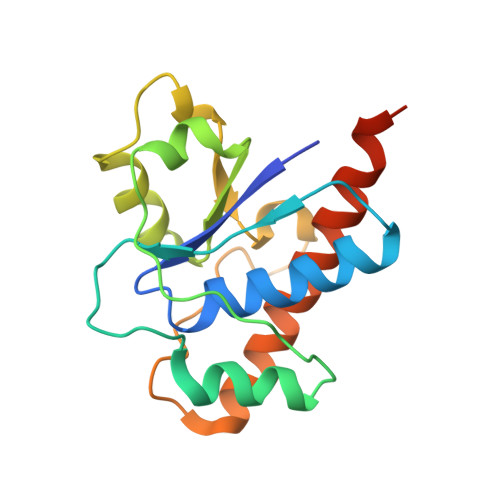
    Low molecular weight protein tyrosine phosphatases (LMWPTPs) are ubiquitously found as small cytoplasmic enzymes which act on phospho-tyrosine containing proteins that are engaged in various cellular functions. Vibrio cholerae O395 contains two LMWPTPs having widely different sequence. Phylogenetic analysis based on a non redundant set of 124 LMWPTP sequences, designate that LMWPTP-2 from Vibrio choleraeO395 (VcLMWPTP-2) is a single taxon. We have determined the crystal structure of VcLMWPTP-2 at 2.6 Å with MOPS bound in the active site. Tertiary structure analysis indicates that VcLMWPTP-2 forms dimer. Studies in solution state also confirm exclusive presence of a dimeric form. Kinetic studies demonstrate that VcLMWPTP-2 dimer is catalytically active while inactivation through oligomerisation was reported as one of the regulatory mechanism in case of mammalian LMWPTP viz., Bos taurus LMWPTP, BPTP. Kinetic studies using p-nitrophenyl phosphate (p-NPP) as a substrate demonstrate active participation of both the P-loop cysteine in catalysis. Vicinal Cys17, in addition plays a role of protecting the catalytic Cys12 under oxidative stress. Structural analysis and MD simulations allowed us to propose the role of several conserved residues around the active site. Distribution of surface charges and grooves around the active site delineates unique features of VcLMWPTP-2 which could be utilized to design specific inhibitor.


  • Organizational Affiliation

    Crystallography and Molecular Biology Division, Saha Institute of Nuclear Physics, HBNI, 1/AF Bidhan Nagar, Kolkata 700064, India.


Macromolecules
Find similar proteins by:  (by identity cutoff)  |  3D Structure
Entity ID: 1
MoleculeChains Sequence LengthOrganismDetailsImage
Phosphotyrosine protein phosphatase
A, B
166Vibrio cholerae O395Mutation(s): 0 
Gene Names: VC0395_A0440
EC: 3.1.3.48
UniProt
Find proteins for A0A0H3AM12 (Vibrio cholerae serotype O1 (strain ATCC 39541 / Classical Ogawa 395 / O395))
Explore A0A0H3AM12 
Go to UniProtKB:  A0A0H3AM12
Entity Groups  
Sequence Clusters30% Identity50% Identity70% Identity90% Identity95% Identity100% Identity
UniProt GroupA0A0H3AM12
Sequence Annotations
Expand
  • Reference Sequence
Small Molecules
Ligands 3 Unique
IDChains Name / Formula / InChI Key2D Diagram3D Interactions
MPO
Query on MPO

Download Ideal Coordinates CCD File 
AA [auth B],
C [auth A]
3[N-MORPHOLINO]PROPANE SULFONIC ACID
C7 H15 N O4 S
DVLFYONBTKHTER-UHFFFAOYSA-N
SO4
Query on SO4

Download Ideal Coordinates CCD File 
BA [auth B],
D [auth A]
SULFATE ION
O4 S
QAOWNCQODCNURD-UHFFFAOYSA-L
GOL
Query on GOL

Download Ideal Coordinates CCD File 
CA [auth B]
DA [auth B]
E [auth A]
EA [auth B]
F [auth A]
CA [auth B],
DA [auth B],
E [auth A],
EA [auth B],
F [auth A],
FA [auth B],
G [auth A],
GA [auth B],
H [auth A],
HA [auth B],
I [auth A],
IA [auth B],
J [auth A],
JA [auth B],
K [auth A],
KA [auth B],
L [auth A],
LA [auth B],
M [auth A],
MA [auth B],
N [auth A],
NA [auth B],
O [auth A],
OA [auth B],
P [auth A],
Q [auth A],
R [auth A],
S [auth A],
T [auth A],
U [auth A],
V [auth A],
W [auth A],
X [auth A],
Y [auth A],
Z [auth A]
GLYCEROL
C3 H8 O3
PEDCQBHIVMGVHV-UHFFFAOYSA-N
Experimental Data & Validation

Experimental Data

  • Method: X-RAY DIFFRACTION
  • Resolution: 2.60 Å
  • R-Value Free: 0.288 
  • R-Value Work: 0.252 
  • R-Value Observed: 0.254 
  • Space Group: C 1 2 1
Unit Cell:
Length ( Å )Angle ( ˚ )
a = 125.912α = 90
b = 51.867β = 103.62
c = 83.896γ = 90
Software Package:
Software NamePurpose
PHENIXrefinement
XDSdata reduction
Aimlessdata scaling
MOLREPphasing

Structure Validation

View Full Validation Report


Ligand Structure Quality Assessment 


Entry History 

Deposition Data

Revision History  (Full details and data files)

  • Version 1.0: 2018-09-19
    Type: Initial release
  • Version 1.1: 2020-05-13
    Changes: Database references
  • Version 1.2: 2023-11-22
    Changes: Data collection, Database references, Refinement description