5Z2W

Crystal structure of the bacterial cell division protein FtsQ and FtsB


Experimental Data Snapshot

  • Method: X-RAY DIFFRACTION
  • Resolution: 3.00 Å
  • R-Value Free: 0.275 
  • R-Value Work: 0.207 
  • R-Value Observed: 0.214 

wwPDB Validation   3D Report Full Report


This is version 1.2 of the entry. See complete history


Literature

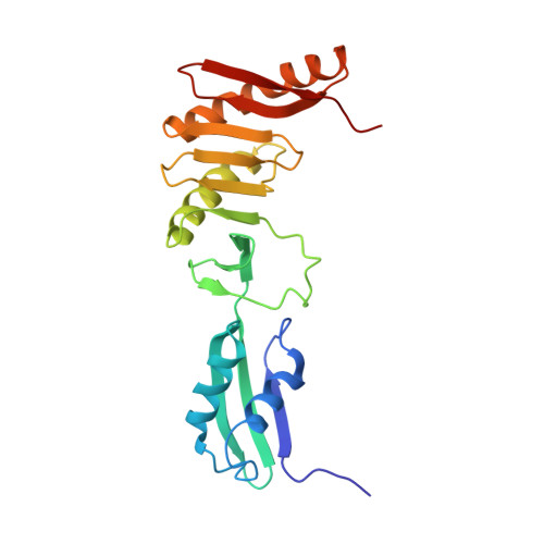
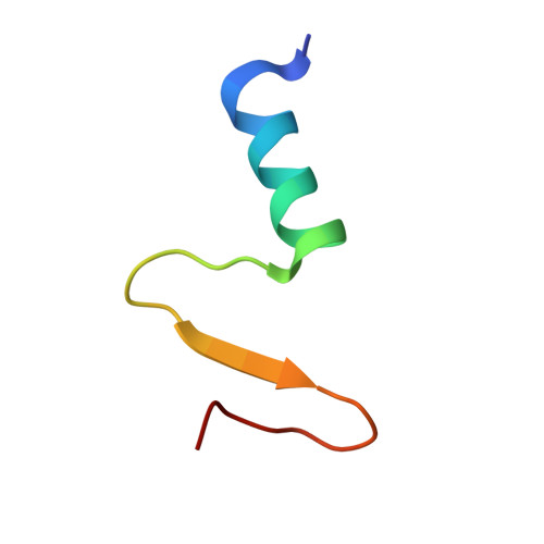
Structural Insights into the FtsQ/FtsB/FtsL Complex, a Key Component of the Divisome.

Choi, Y.Kim, J.Yoon, H.J.Jin, K.S.Ryu, S.Lee, H.H.

(2018) Sci Rep 8: 18061-18061

  • DOI: https://doi.org/10.1038/s41598-018-36001-2
  • Primary Citation of Related Structures:  
    5Z2W

  • PubMed Abstract: 

    Bacterial cell division is a fundamental process that results in the physical separation of a mother cell into two daughter cells and involves a set of proteins known as the divisome. Among them, the FtsQ/FtsB/FtsL complex was known as a scaffold protein complex, but its overall structure and exact function is not precisely known. In this study, we have determined the crystal structure of the periplasmic domain of FtsQ in complex with the C-terminal fragment of FtsB, and showed that the C-terminal region of FtsB is a key binding region of FtsQ via mutational analysis in vitro and in vivo. We also obtained the solution structure of the periplasmic FtsQ/FtsB/FtsL complex by small angle X-ray scattering (SAXS), which reveals its structural organization. Interestingly, the SAXS and analytical gel filtration data showed that the FtsQ/FtsB/FtsL complex forms a 2:2:2 heterohexameric assembly in solution with the "Y" shape. Based on the model, the N-terminal directions of FtsQ and the FtsB/FtsL complex should be opposite, suggesting that the Y-shaped FtsQ/FtsB/FtsL complex might fit well into the curved membrane for membrane anchoring.


  • Organizational Affiliation

    Department of Chemistry, College of Natural Sciences, Seoul National University, Seoul, 08826, Korea.


Macromolecules
Find similar proteins by:  (by identity cutoff)  |  3D Structure
Entity ID: 1
MoleculeChains Sequence LengthOrganismDetailsImage
Cell division protein FtsQ209Escherichia coliMutation(s): 0 
Gene Names: ftsQ
UniProt
Find proteins for P06136 (Escherichia coli (strain K12))
Explore P06136 
Go to UniProtKB:  P06136
Entity Groups  
Sequence Clusters30% Identity50% Identity70% Identity90% Identity95% Identity100% Identity
UniProt GroupP06136
Sequence Annotations
Expand
  • Reference Sequence
Find similar proteins by:  (by identity cutoff)  |  3D Structure
Entity ID: 2
MoleculeChains Sequence LengthOrganismDetailsImage
Cell division protein FtsB35Escherichia coliMutation(s): 0 
Gene Names: ftsBAWP75_20790
UniProt
Find proteins for P0A6S5 (Escherichia coli (strain K12))
Explore P0A6S5 
Go to UniProtKB:  P0A6S5
Entity Groups  
Sequence Clusters30% Identity50% Identity70% Identity90% Identity95% Identity100% Identity
UniProt GroupP0A6S5
Sequence Annotations
Expand
  • Reference Sequence
Experimental Data & Validation

Experimental Data

  • Method: X-RAY DIFFRACTION
  • Resolution: 3.00 Å
  • R-Value Free: 0.275 
  • R-Value Work: 0.207 
  • R-Value Observed: 0.214 
  • Space Group: P 1 2 1
Unit Cell:
Length ( Å )Angle ( ˚ )
a = 63.557α = 90
b = 39.306β = 94.77
c = 73.191γ = 90
Software Package:
Software NamePurpose
REFMACrefinement
HKL-2000data reduction
HKL-2000data scaling
MOLREPphasing
PDB_EXTRACTdata extraction

Structure Validation

View Full Validation Report


Entry History 

Deposition Data

Revision History  (Full details and data files)

  • Version 1.0: 2019-01-02
    Type: Initial release
  • Version 1.1: 2019-01-16
    Changes: Data collection, Database references
  • Version 1.2: 2024-03-27
    Changes: Data collection, Database references