5Z2V

Crystal structure of RecR from Pseudomonas aeruginosa PAO1


Experimental Data Snapshot

  • Method: X-RAY DIFFRACTION
  • Resolution: 2.20 Å
  • R-Value Free: 0.213 
  • R-Value Work: 0.178 
  • R-Value Observed: 0.180 

wwPDB Validation   3D Report Full Report


This is version 1.1 of the entry. See complete history


Literature

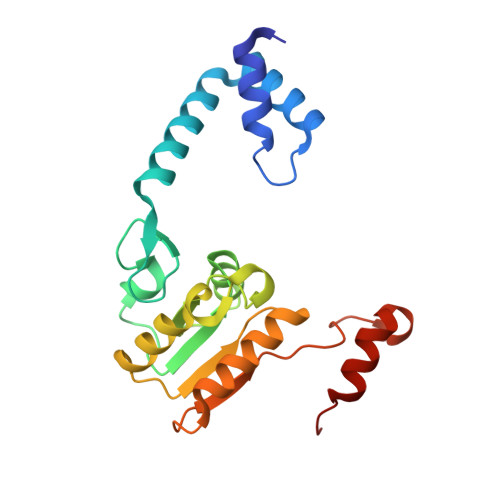
Crystal structure of RecR, a member of the RecFOR DNA-repair pathway, from Pseudomonas aeruginosa PAO1.

Che, S.Chen, Y.Liang, Y.Zhang, Q.Bartlam, M.

(2018) Acta Crystallogr F Struct Biol Commun 74: 222-230

  • DOI: https://doi.org/10.1107/S2053230X18003503
  • Primary Citation of Related Structures:  
    5Z2V

  • PubMed Abstract: 

    DNA damage is usually lethal to all organisms. Homologous recombination plays an important role in the DNA damage-repair process in prokaryotic organisms. Two pathways are responsible for homologous recombination in Pseudomonas aeruginosa: the RecBCD pathway and the RecFOR pathway. RecR is an important regulator in the RecFOR homologous recombination pathway in P. aeruginosa. It forms complexes with RecF and RecO that can facilitate the loading of RecA onto ssDNA in the RecFOR pathway. Here, the crystal structure of RecR from P. aeruginosa PAO1 (PaRecR) is reported. PaRecR crystallizes in space group P6 1 22, with two monomers per asymmetric unit. Analytical ultracentrifugation data show that PaRecR forms a stable dimer, but can exist as a tetramer in solution. The crystal structure shows that dimeric PaRecR forms a ring-like tetramer architecture via crystal symmetry. The presence of a ligand in the Walker B motif of one RecR subunit suggests a putative nucleotide-binding site.


  • Organizational Affiliation

    College of Life Sciences, Nankai University, 94 Weijin Road, Tianjin 300071, People's Republic of China.


Macromolecules
Find similar proteins by:  (by identity cutoff)  |  3D Structure
Entity ID: 1
MoleculeChains Sequence LengthOrganismDetailsImage
Recombination protein RecR
A, B
198Pseudomonas aeruginosa PAO1Mutation(s): 0 
Gene Names: recRPA1534
UniProt
Find proteins for Q9I3H9 (Pseudomonas aeruginosa (strain ATCC 15692 / DSM 22644 / CIP 104116 / JCM 14847 / LMG 12228 / 1C / PRS 101 / PAO1))
Explore Q9I3H9 
Go to UniProtKB:  Q9I3H9
Entity Groups  
Sequence Clusters30% Identity50% Identity70% Identity90% Identity95% Identity100% Identity
UniProt GroupQ9I3H9
Sequence Annotations
Expand
  • Reference Sequence
Small Molecules
Ligands 5 Unique
IDChains Name / Formula / InChI Key2D Diagram3D Interactions
N
Query on N

Download Ideal Coordinates CCD File 
F [auth A]ANY 5'-MONOPHOSPHATE NUCLEOTIDE
C5 H11 O7 P
CYZZKTRFOOKUMT-LMVFSUKVSA-N
PO4
Query on PO4

Download Ideal Coordinates CCD File 
H [auth B]PHOSPHATE ION
O4 P
NBIIXXVUZAFLBC-UHFFFAOYSA-K
GOL
Query on GOL

Download Ideal Coordinates CCD File 
D [auth A],
E [auth A]
GLYCEROL
C3 H8 O3
PEDCQBHIVMGVHV-UHFFFAOYSA-N
ZN
Query on ZN

Download Ideal Coordinates CCD File 
C [auth A],
G [auth B]
ZINC ION
Zn
PTFCDOFLOPIGGS-UHFFFAOYSA-N
NA
Query on NA

Download Ideal Coordinates CCD File 
I [auth B]SODIUM ION
Na
FKNQFGJONOIPTF-UHFFFAOYSA-N
Experimental Data & Validation

Experimental Data

  • Method: X-RAY DIFFRACTION
  • Resolution: 2.20 Å
  • R-Value Free: 0.213 
  • R-Value Work: 0.178 
  • R-Value Observed: 0.180 
  • Space Group: P 61 2 2
Unit Cell:
Length ( Å )Angle ( ˚ )
a = 70.075α = 90
b = 70.075β = 90
c = 369.022γ = 120
Software Package:
Software NamePurpose
REFMACrefinement
HKL-2000data reduction
HKL-2000data scaling
PHASERphasing

Structure Validation

View Full Validation Report


Entry History & Funding Information

Deposition Data


Funding OrganizationLocationGrant Number
National Natural Science Foundation of ChinaChina31570128
Tianjin Municipal Science and Technology CommissionChina13JCYBJC20800

Revision History  (Full details and data files)

  • Version 1.0: 2019-01-02
    Type: Initial release
  • Version 1.1: 2023-11-22
    Changes: Data collection, Database references, Derived calculations, Refinement description