5Z1W

Cryo-EM structure of polycystic kidney disease-like channel PKD2L1


Experimental Data Snapshot

  • Method: ELECTRON MICROSCOPY
  • Resolution: 3.38 Å
  • Aggregation State: PARTICLE 
  • Reconstruction Method: SINGLE PARTICLE 

wwPDB Validation   3D Report Full Report


This is version 1.2 of the entry. See complete history


Literature

Cryo-EM structure of the polycystic kidney disease-like channel PKD2L1.

Su, Q.Hu, F.Liu, Y.Ge, X.Mei, C.Yu, S.Shen, A.Zhou, Q.Yan, C.Lei, J.Zhang, Y.Liu, X.Wang, T.

(2018) Nat Commun 9: 1192-1192

  • DOI: https://doi.org/10.1038/s41467-018-03606-0
  • Primary Citation of Related Structures:  
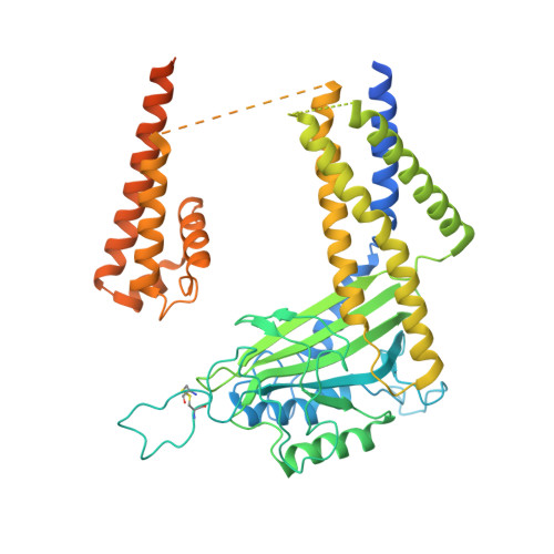
    5Z1W

  • PubMed Abstract: 

    PKD2L1, also termed TRPP3 from the TRPP subfamily (polycystic TRP channels), is involved in the sour sensation and other pH-dependent processes. PKD2L1 is believed to be a nonselective cation channel that can be regulated by voltage, protons, and calcium. Despite its considerable importance, the molecular mechanisms underlying PKD2L1 regulations are largely unknown. Here, we determine the PKD2L1 atomic structure at 3.38 Å resolution by cryo-electron microscopy, whereby side chains of nearly all residues are assigned. Unlike its ortholog PKD2, the pore helix (PH) and transmembrane segment 6 (S6) of PKD2L1, which are involved in upper and lower-gate opening, adopt an open conformation. Structural comparisons of PKD2L1 with a PKD2-based homologous model indicate that the pore domain dilation is coupled to conformational changes of voltage-sensing domains (VSDs) via a series of π-π interactions, suggesting a potential PKD2L1 gating mechanism.


  • Organizational Affiliation

    Ministry of Education Key Laboratory of Protein Science, Tsinghua University, Beijing, 100084, China.


Macromolecules
Find similar proteins by:  (by identity cutoff)  |  3D Structure
Entity ID: 1
MoleculeChains Sequence LengthOrganismDetailsImage
Polycystic kidney disease 2-like 1 protein
A, B, C, D
566Mus musculusMutation(s): 0 
Gene Names: Pkd2l1Trpp3
Membrane Entity: Yes 
UniProt
Find proteins for A2A259 (Mus musculus)
Explore A2A259 
Go to UniProtKB:  A2A259
Entity Groups  
Sequence Clusters30% Identity50% Identity70% Identity90% Identity95% Identity100% Identity
UniProt GroupA2A259
Sequence Annotations
Expand
  • Reference Sequence
Small Molecules
Ligands 1 Unique
IDChains Name / Formula / InChI Key2D Diagram3D Interactions
NAG
Query on NAG

Download Ideal Coordinates CCD File 
E [auth A]
F [auth A]
G [auth A]
H [auth B]
I [auth B]
E [auth A],
F [auth A],
G [auth A],
H [auth B],
I [auth B],
J [auth B],
K [auth C],
L [auth C],
M [auth C],
N [auth D],
O [auth D],
P [auth D]
2-acetamido-2-deoxy-beta-D-glucopyranose
C8 H15 N O6
OVRNDRQMDRJTHS-FMDGEEDCSA-N
Experimental Data & Validation

Experimental Data

  • Method: ELECTRON MICROSCOPY
  • Resolution: 3.38 Å
  • Aggregation State: PARTICLE 
  • Reconstruction Method: SINGLE PARTICLE 

Structure Validation

View Full Validation Report


Entry History & Funding Information

Deposition Data

  • Released Date: 2018-03-28 
  • Deposition Author(s): Zhang, Y.Q.

Funding OrganizationLocationGrant Number
China--

Revision History  (Full details and data files)

  • Version 1.0: 2018-03-28
    Type: Initial release
  • Version 1.1: 2019-04-17
    Changes: Data collection, Database references
  • Version 1.2: 2020-07-29
    Type: Remediation
    Reason: Carbohydrate remediation
    Changes: Data collection, Derived calculations, Structure summary