5Z10

Structure of the mechanosensitive Piezo1 channel


Experimental Data Snapshot

  • Method: ELECTRON MICROSCOPY
  • Resolution: 3.97 Å
  • Aggregation State: PARTICLE 
  • Reconstruction Method: SINGLE PARTICLE 

wwPDB Validation   3D Report Full Report


This is version 2.0 of the entry. See complete history


Literature

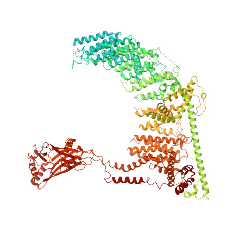
Structure and mechanogating mechanism of the Piezo1 channel.

Zhao, Q.Zhou, H.Chi, S.Wang, Y.Wang, J.Geng, J.Wu, K.Liu, W.Zhang, T.Dong, M.Q.Wang, J.Li, X.Xiao, B.

(2018) Nature 554: 487-492

  • DOI: https://doi.org/10.1038/nature25743
  • Primary Citation of Related Structures:  
    5Z10

  • PubMed Abstract: 

    The mechanosensitive Piezo channels function as key eukaryotic mechanotransducers. However, their structures and mechanogating mechanisms remain unknown. Here we determine the three-bladed, propeller-like electron cryo-microscopy structure of mouse Piezo1 and functionally reveal its mechanotransduction components. Despite the lack of sequence repetition, we identify nine repetitive units consisting of four transmembrane helices each-which we term transmembrane helical units (THUs)-which assemble into a highly curved blade-like structure. The last transmembrane helix encloses a hydrophobic pore, followed by three intracellular fenestration sites and side portals that contain pore-property-determining residues. The central region forms a 90 Å-long intracellular beam-like structure, which undergoes a lever-like motion to connect THUs to the pore via the interfaces of the C-terminal domain, the anchor-resembling domain and the outer helix. Deleting extracellular loops in the distal THUs or mutating single residues in the beam impairs the mechanical activation of Piezo1. Overall, Piezo1 possesses a unique 38-transmembrane-helix topology and designated mechanotransduction components, which enable a lever-like mechanogating mechanism.


  • Organizational Affiliation

    School of Pharmaceutical Sciences or Life Sciences, Tsinghua University, Beijing 100084, China.


Macromolecules
Find similar proteins by:  (by identity cutoff)  |  3D Structure
Entity ID: 1
MoleculeChains Sequence LengthOrganismDetailsImage
Piezo-type mechanosensitive ion channel component 1
A, B, C
2,547Mus musculusMutation(s): 0 
Gene Names: Piezo1Fam38a
Membrane Entity: Yes 
UniProt & NIH Common Fund Data Resources
Find proteins for E2JF22 (Mus musculus)
Explore E2JF22 
Go to UniProtKB:  E2JF22
IMPC:  MGI:3603204
Entity Groups  
Sequence Clusters30% Identity50% Identity70% Identity90% Identity95% Identity100% Identity
UniProt GroupE2JF22
Sequence Annotations
Expand
  • Reference Sequence
Experimental Data & Validation

Experimental Data

  • Method: ELECTRON MICROSCOPY
  • Resolution: 3.97 Å
  • Aggregation State: PARTICLE 
  • Reconstruction Method: SINGLE PARTICLE 
EM Software:
TaskSoftware PackageVersion
RECONSTRUCTIONRELION1.4

Structure Validation

View Full Validation Report


Entry History & Funding Information

Deposition Data


Funding OrganizationLocationGrant Number
National Natural Science Foundation of China (NSFC)China31630090

Revision History  (Full details and data files)

  • Version 1.0: 2018-01-31
    Type: Initial release
  • Version 1.1: 2018-02-07
    Changes: Database references, Structure summary
  • Version 1.2: 2018-03-07
    Changes: Database references
  • Version 2.0: 2020-01-29
    Type: Coordinate replacement
    Reason: Model completeness
    Changes: Advisory, Atomic model, Author supporting evidence, Data collection, Derived calculations, Refinement description, Structure summary