5Z06

Crystal structure of beta-1,2-glucanase from Parabacteroides distasonis


Experimental Data Snapshot

  • Method: X-RAY DIFFRACTION
  • Resolution: 2.10 Å
  • R-Value Free: 0.219 
  • R-Value Work: 0.178 
  • R-Value Observed: 0.180 

wwPDB Validation   3D Report Full Report


This is version 1.2 of the entry. See complete history


Literature

Characterization and Structural Analysis of a Novel exo-Type Enzyme Acting on beta-1,2-Glucooligosaccharides from Parabacteroides distasonis

Shimizu, H.Nakajima, M.Miyanaga, A.Takahashi, Y.Tanaka, N.Kobayashi, K.Sugimoto, N.Nakai, H.Taguchi, H.

(2018) Biochemistry 57: 3849-3860

  • DOI: https://doi.org/10.1021/acs.biochem.8b00385
  • Primary Citation of Related Structures:  
    5Z06

  • PubMed Abstract: 

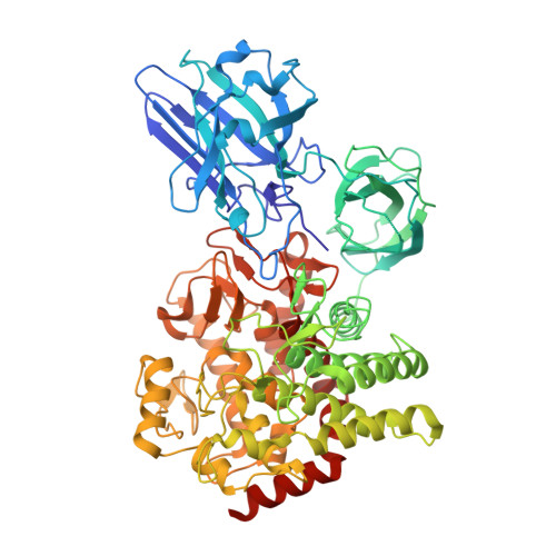
    β-1,2-Glucan is a polysaccharide produced mainly by some Gram-negative bacteria as a symbiosis and infectious factor. We recently identified endo-β-1,2-glucanase from Chitinophaga pinensis ( CpSGL) as an enzyme comprising a new family. Here, we report the characteristics and crystal structure of a CpSGL homologue from Parabacteroides distasonis, an intestinal bacterium (BDI_3064 protein), which exhibits distinctive properties of known β-1,2-glucan-degrading enzymes. BDI_3064 hydrolyzed linear β-1,2-glucan and β-1,2-glucooligosaccharides with degrees of polymerization (DPs) of ≥4 to produce sophorose specifically but did not hydrolyze cyclic β-1,2-glucan. This result indicates that BDI_3064 is a new exo-type enzyme. BDI_3064 also produced sophorose from β-1,2-glucooligosaccharide analogues that have a modified reducing end, indicating that BDI_3064 acts on its substrates from the nonreducing end. The crystal structure showed that BDI_3064 possesses additional N-terminal domains 1 and 2, unlike CpSGL. Superimposition of BDI_3064 and CpSGL complexed with ligands showed that R93 in domain 1 overlapped subsite -3 in CpSGL. Docking analysis involving a β-1,2-glucooligosaccharide with DP4 showed that R93 completely blocks the nonreducing end of the docked β-1,2-glucooligosaccharide. This indicates that BDI_3064 employs a distinct mechanism of recognition at the nonreducing end of substrates to act as an exo-type enzyme. Thus, we propose 2-β-d-glucooligosaccharide sophorohydrolase (nonreducing end) as a systematic name for BDI_3064.


  • Organizational Affiliation

    Department of Applied Biological Science, Faculty of Science and Technology , Tokyo University of Science , 2641 Yamazaki , Noda , Chiba 278-8510 , Japan.


Macromolecules
Find similar proteins by:  (by identity cutoff)  |  3D Structure
Entity ID: 1
MoleculeChains Sequence LengthOrganismDetailsImage
BDI_3064 protein
A, B
712Parabacteroides distasonis ATCC 8503Mutation(s): 0 
Gene Names: BDI_3064
UniProt
Find proteins for A6LGF6 (Parabacteroides distasonis (strain ATCC 8503 / DSM 20701 / CIP 104284 / JCM 5825 / NCTC 11152))
Explore A6LGF6 
Go to UniProtKB:  A6LGF6
Entity Groups  
Sequence Clusters30% Identity50% Identity70% Identity90% Identity95% Identity100% Identity
UniProt GroupA6LGF6
Sequence Annotations
Expand
  • Reference Sequence
Small Molecules
Ligands 2 Unique
IDChains Name / Formula / InChI Key2D Diagram3D Interactions
GOL
Query on GOL

Download Ideal Coordinates CCD File 
D [auth A]
E [auth A]
F [auth A]
G [auth A]
H [auth A]
D [auth A],
E [auth A],
F [auth A],
G [auth A],
H [auth A],
I [auth A],
K [auth B],
L [auth B],
M [auth B],
N [auth B]
GLYCEROL
C3 H8 O3
PEDCQBHIVMGVHV-UHFFFAOYSA-N
CA
Query on CA

Download Ideal Coordinates CCD File 
C [auth A],
J [auth B]
CALCIUM ION
Ca
BHPQYMZQTOCNFJ-UHFFFAOYSA-N
Experimental Data & Validation

Experimental Data

  • Method: X-RAY DIFFRACTION
  • Resolution: 2.10 Å
  • R-Value Free: 0.219 
  • R-Value Work: 0.178 
  • R-Value Observed: 0.180 
  • Space Group: C 1 2 1
Unit Cell:
Length ( Å )Angle ( ˚ )
a = 130.08α = 90
b = 123.337β = 107.88
c = 101.376γ = 90
Software Package:
Software NamePurpose
REFMACrefinement
XDSdata reduction
Aimlessdata scaling
BUCCANEERmodel building
MOLREPphasing

Structure Validation

View Full Validation Report


Entry History & Funding Information

Deposition Data


Funding OrganizationLocationGrant Number
Mishima Kaiun Memorial FoundationJapan--

Revision History  (Full details and data files)

  • Version 1.0: 2018-05-30
    Type: Initial release
  • Version 1.1: 2018-07-18
    Changes: Data collection, Database references, Source and taxonomy
  • Version 1.2: 2023-11-22
    Changes: Data collection, Database references, Refinement description