5YW2

Crystal structure of Adenine phosphoribosyltransferase from Francisella tularensis.


Experimental Data Snapshot

  • Method: X-RAY DIFFRACTION
  • Resolution: 2.28 Å
  • R-Value Free: 0.259 
  • R-Value Work: 0.202 
  • R-Value Observed: 0.205 

wwPDB Validation   3D Report Full Report


This is version 1.3 of the entry. See complete history


Literature

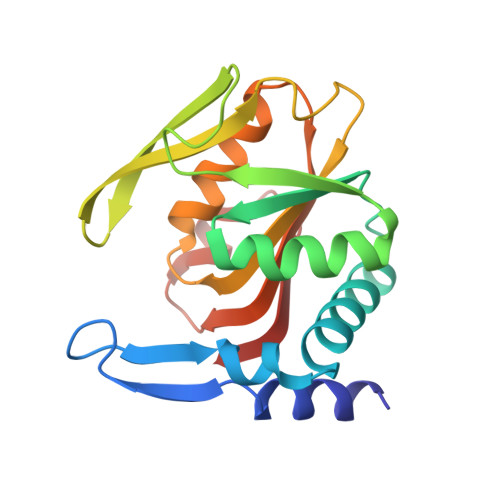
Crystal structures of APRT from Francisella tularensis - an N-H···N hydrogen bond imparts adenine specificity in adenine phosporibosyltransferases.

Pavithra, G.C.Ramagopal, U.A.

(2018) FEBS J 285: 2306-2318

  • DOI: https://doi.org/10.1111/febs.14481
  • Primary Citation of Related Structures:  
    5YW2, 5YW5

  • PubMed Abstract: 

    Francisella tularensisis, the causative agent of tularemia has been classified as a category A bioterrorism agent. Here, we present the crystal structure of apo and adenine bound form of the adenine phosphoribosyltransferase (APRT) from Francisella tularensis. APRT is an enzyme involved in the salvage of adenine (a 6-aminopurine), converting it to AMP. The purine salvage pathway relies on two essential and distinct enzymes to convert 6-aminopurine and 6-oxopurines into corresponding nucleotides. The mechanism by which these enzymes differentiate different purines is not clearly understood. Analysis of the structures of apo and adenine-bound APRT from F. tularensis, together with all other available structures of APRTs, suggests that (a) the base-binding loop is stabilized by a cluster of aromatic and conformation-restricting proline residues, and (b) an N-H···N hydrogen bond between the base-binding loop and the N1 atom of adenine is the key interaction that differentiates adenine from 6-oxopurines. These observations were corroborated by bioinformatics analysis of ~ 4000 sequences of APRTs (with 80% identity cutoff), which confirmed that the residues conferring rigidity to the base-binding loop are highly conserved. Furthermore, an F23A mutation on the base-binding loop severely affects the efficiency of the enzyme. We extended our analysis to the structure and sequences of APRTs from the Trypanosomatidae family with a destabilizing insertion on the base-binding loop and propose the mechanism by which these evolutionarily divergent enzymes achieve base specificity. Our results suggest that the base-binding loop not only confers appropriate affinity but also provides defined specificity for adenine.


  • Organizational Affiliation

    Division of Biological Sciences, Poornaprajna Institute of Scientific Research, Bangalore, India.


Macromolecules
Find similar proteins by:  (by identity cutoff)  |  3D Structure
Entity ID: 1
MoleculeChains Sequence LengthOrganismDetailsImage
Adenine phosphoribosyltransferaseA,
B [auth C],
C [auth B],
D
179Francisella tularensisMutation(s): 0 
Gene Names: apt
EC: 2.4.2.7
UniProt
Find proteins for Q5NII9 (Francisella tularensis subsp. tularensis (strain SCHU S4 / Schu 4))
Explore Q5NII9 
Go to UniProtKB:  Q5NII9
Entity Groups  
Sequence Clusters30% Identity50% Identity70% Identity90% Identity95% Identity100% Identity
UniProt GroupQ5NII9
Sequence Annotations
Expand
  • Reference Sequence
Small Molecules
Ligands 3 Unique
IDChains Name / Formula / InChI Key2D Diagram3D Interactions
SO4
Query on SO4

Download Ideal Coordinates CCD File 
E [auth A],
K [auth C],
N [auth B],
P [auth D]
SULFATE ION
O4 S
QAOWNCQODCNURD-UHFFFAOYSA-L
CL
Query on CL

Download Ideal Coordinates CCD File 
G [auth A]
H [auth A]
I [auth A]
J [auth A]
L [auth C]
G [auth A],
H [auth A],
I [auth A],
J [auth A],
L [auth C],
M [auth C],
O [auth D],
R [auth D]
CHLORIDE ION
Cl
VEXZGXHMUGYJMC-UHFFFAOYSA-M
MG
Query on MG

Download Ideal Coordinates CCD File 
F [auth A],
Q [auth D]
MAGNESIUM ION
Mg
JLVVSXFLKOJNIY-UHFFFAOYSA-N
Experimental Data & Validation

Experimental Data

  • Method: X-RAY DIFFRACTION
  • Resolution: 2.28 Å
  • R-Value Free: 0.259 
  • R-Value Work: 0.202 
  • R-Value Observed: 0.205 
  • Space Group: P 21 21 21
Unit Cell:
Length ( Å )Angle ( ˚ )
a = 54.168α = 90
b = 82.147β = 90
c = 170.119γ = 90
Software Package:
Software NamePurpose
REFMACrefinement
SCALEPACKdata scaling
PDB_EXTRACTdata extraction
DENZOdata reduction
MOLREPphasing

Structure Validation

View Full Validation Report


Entry History 

Deposition Data

Revision History  (Full details and data files)

  • Version 1.0: 2018-01-03
    Type: Initial release
  • Version 1.1: 2018-05-09
    Changes: Data collection, Database references
  • Version 1.2: 2018-07-04
    Changes: Data collection, Database references
  • Version 1.3: 2023-11-22
    Changes: Data collection, Database references, Derived calculations, Refinement description