5YVQ

Complex of Mu phage tail fiber and its chaperone


Experimental Data Snapshot

  • Method: X-RAY DIFFRACTION
  • Resolution: 2.10 Å
  • R-Value Free: 0.264 
  • R-Value Work: 0.236 
  • R-Value Observed: 0.238 

wwPDB Validation   3D Report Full Report


This is version 1.2 of the entry. See complete history


Literature

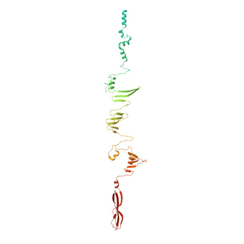
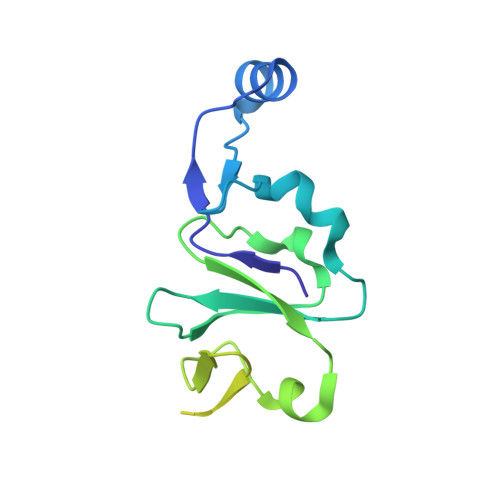
Phage tail fibre assembly proteins employ a modular structure to drive the correct folding of diverse fibres.

North, O.I.Sakai, K.Yamashita, E.Nakagawa, A.Iwazaki, T.Buttner, C.R.Takeda, S.Davidson, A.R.

(2019) Nat Microbiol 4: 1645-1653

  • DOI: https://doi.org/10.1038/s41564-019-0477-7
  • Primary Citation of Related Structures:  
    5YVQ

  • PubMed Abstract: 

    Phage tail fibres are elongated protein assemblies capable of specific recognition of bacterial surfaces during the first step of viral infection 1-4 . The folding of these complex trimeric structures often requires a phage-encoded tail fibre assembly (Tfa) protein 5-7 . Despite the wide occurrence of Tfa proteins, their functional mechanism has not been elucidated. Here, we investigate the tail fibre and Tfa of Escherichia coli phage Mu. We demonstrate that Tfa forms a stable complex with the tail fibre, and present a 2.1 Å resolution X-ray crystal structure of this complex. We find that Tfa proteins are comprised of two domains: a non-conserved N-terminal domain that binds to the C-terminal region of the fibre and a conserved C-terminal domain that probably mediates fibre oligomerization and assembly. Tfa forms rapidly exchanging multimers on its own, but not a stable trimer, implying that Tfa does not specify the trimeric state of the fibre. We propose that the key conserved role of Tfa is to ensure that fibre assembly and multimerization initiates at the C terminus, ensuring that the intertwined and repetitive structural elements of fibres come together in the correct sequence. The universal importance of correctly aligning the C termini of phage fibres is highlighted by our work.


  • Organizational Affiliation

    Department of Biochemistry, University of Toronto, Toronto, Ontario, Canada.


Macromolecules
Find similar proteins by:  (by identity cutoff)  |  3D Structure
Entity ID: 1
MoleculeChains Sequence LengthOrganismDetailsImage
Tail fiber protein S504Muvirus muMutation(s): 0 
Gene Names: SMup49
UniProt
Find proteins for Q9T1V0 (Escherichia phage Mu)
Explore Q9T1V0 
Go to UniProtKB:  Q9T1V0
Entity Groups  
Sequence Clusters30% Identity50% Identity70% Identity90% Identity95% Identity100% Identity
UniProt GroupQ9T1V0
Sequence Annotations
Expand
  • Reference Sequence
Find similar proteins by:  (by identity cutoff)  |  3D Structure
Entity ID: 2
MoleculeChains Sequence LengthOrganismDetailsImage
Tail fiber assembly protein U175Muvirus muMutation(s): 0 
Gene Names: UMup50
UniProt
Find proteins for Q9T1U9 (Escherichia phage Mu)
Explore Q9T1U9 
Go to UniProtKB:  Q9T1U9
Entity Groups  
Sequence Clusters30% Identity50% Identity70% Identity90% Identity95% Identity100% Identity
UniProt GroupQ9T1U9
Sequence Annotations
Expand
  • Reference Sequence
Experimental Data & Validation

Experimental Data

  • Method: X-RAY DIFFRACTION
  • Resolution: 2.10 Å
  • R-Value Free: 0.264 
  • R-Value Work: 0.236 
  • R-Value Observed: 0.238 
  • Space Group: P 3 2 1
Unit Cell:
Length ( Å )Angle ( ˚ )
a = 66.332α = 90
b = 66.332β = 90
c = 387.769γ = 120
Software Package:
Software NamePurpose
PHENIXrefinement
HKL-2000data reduction
HKL-2000data scaling
SHARPphasing

Structure Validation

View Full Validation Report


Entry History 

Deposition Data

Revision History  (Full details and data files)

  • Version 1.0: 2019-05-22
    Type: Initial release
  • Version 1.1: 2019-10-09
    Changes: Data collection, Database references
  • Version 1.2: 2024-03-27
    Changes: Data collection, Database references