5YVI

Crystal structure of Karyopherin beta2 in complex with FUS(456-526)


Experimental Data Snapshot

  • Method: X-RAY DIFFRACTION
  • Resolution: 2.90 Å
  • R-Value Free: 0.231 
  • R-Value Work: 0.179 
  • R-Value Observed: 0.182 

wwPDB Validation   3D Report Full Report


This is version 1.1 of the entry. See complete history


Literature

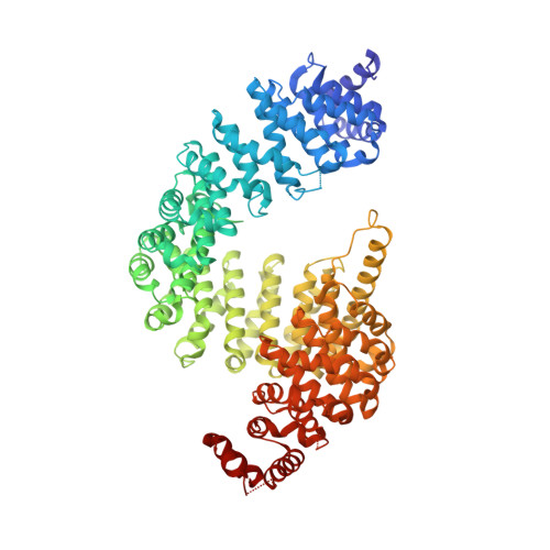
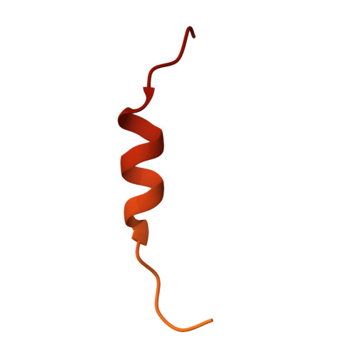
Nuclear Import Receptor Inhibits Phase Separation of FUS through Binding to Multiple Sites.

Yoshizawa, T.Ali, R.Jiou, J.Fung, H.Y.J.Burke, K.A.Kim, S.J.Lin, Y.Peeples, W.B.Saltzberg, D.Soniat, M.Baumhardt, J.M.Oldenbourg, R.Sali, A.Fawzi, N.L.Rosen, M.K.Chook, Y.M.

(2018) Cell 173: 693-705.e22

  • DOI: https://doi.org/10.1016/j.cell.2018.03.003
  • Primary Citation of Related Structures:  
    5YVG, 5YVH, 5YVI

  • PubMed Abstract: 

    Liquid-liquid phase separation (LLPS) is believed to underlie formation of biomolecular condensates, cellular compartments that concentrate macromolecules without surrounding membranes. Physical mechanisms that control condensate formation/dissolution are poorly understood. The RNA-binding protein fused in sarcoma (FUS) undergoes LLPS in vitro and associates with condensates in cells. We show that the importin karyopherin-β2/transportin-1 inhibits LLPS of FUS. This activity depends on tight binding of karyopherin-β2 to the C-terminal proline-tyrosine nuclear localization signal (PY-NLS) of FUS. Nuclear magnetic resonance (NMR) analyses reveal weak interactions of karyopherin-β2 with sequence elements and structural domains distributed throughout the entirety of FUS. Biochemical analyses demonstrate that most of these same regions also contribute to LLPS of FUS. The data lead to a model where high-affinity binding of karyopherin-β2 to the FUS PY-NLS tethers the proteins together, allowing multiple, distributed weak intermolecular contacts to disrupt FUS self-association, blocking LLPS. Karyopherin-β2 may act analogously to control condensates in diverse cellular contexts.


  • Organizational Affiliation

    Department of Pharmacology, University of Texas Southwestern Medical Center, Dallas, TX 75390, USA.


Macromolecules
Find similar proteins by:  (by identity cutoff)  |  3D Structure
Entity ID: 1
MoleculeChains Sequence LengthOrganismDetailsImage
Transportin-1868Homo sapiensMutation(s): 0 
Gene Names: TNPO1KPNB2MIP1TRN
UniProt & NIH Common Fund Data Resources
Find proteins for Q92973 (Homo sapiens)
Explore Q92973 
Go to UniProtKB:  Q92973
PHAROS:  Q92973
GTEx:  ENSG00000083312 
Entity Groups  
Sequence Clusters30% Identity50% Identity70% Identity90% Identity95% Identity100% Identity
UniProt GroupQ92973
Sequence Annotations
Expand
  • Reference Sequence
Find similar proteins by:  (by identity cutoff)  |  3D Structure
Entity ID: 2
MoleculeChains Sequence LengthOrganismDetailsImage
RNA-binding protein FUS73Homo sapiensMutation(s): 0 
Gene Names: FUSTLS
UniProt & NIH Common Fund Data Resources
Find proteins for P35637 (Homo sapiens)
Explore P35637 
Go to UniProtKB:  P35637
PHAROS:  P35637
GTEx:  ENSG00000089280 
Entity Groups  
Sequence Clusters30% Identity50% Identity70% Identity90% Identity95% Identity100% Identity
UniProt GroupP35637
Sequence Annotations
Expand
  • Reference Sequence
Experimental Data & Validation

Experimental Data

  • Method: X-RAY DIFFRACTION
  • Resolution: 2.90 Å
  • R-Value Free: 0.231 
  • R-Value Work: 0.179 
  • R-Value Observed: 0.182 
  • Space Group: P 21 21 2
Unit Cell:
Length ( Å )Angle ( ˚ )
a = 129.042α = 90
b = 156.102β = 90
c = 68.248γ = 90
Software Package:
Software NamePurpose
PHENIXrefinement
HKL-3000data scaling
PHASESphasing

Structure Validation

View Full Validation Report


Entry History 

Deposition Data

Revision History  (Full details and data files)

  • Version 1.0: 2018-05-16
    Type: Initial release
  • Version 1.1: 2023-11-22
    Changes: Data collection, Database references, Refinement description