5YQR

Crystal structure of the PH-like domain of Lam6


Experimental Data Snapshot

  • Method: X-RAY DIFFRACTION
  • Resolution: 2.40 Å
  • R-Value Free: 0.266 
  • R-Value Work: 0.218 
  • R-Value Observed: 0.221 

wwPDB Validation   3D Report Full Report


This is version 1.2 of the entry. See complete history


Literature

Structural basis of sterol recognition and nonvesicular transport by lipid transfer proteins anchored at membrane contact sites

Tong, J.Manik, M.K.Im, Y.J.

(2018) Proc Natl Acad Sci U S A 115: E856-E865

  • DOI: https://doi.org/10.1073/pnas.1719709115
  • Primary Citation of Related Structures:  
    5YQI, 5YQJ, 5YQP, 5YQQ, 5YQR, 5YS0

  • PubMed Abstract: 

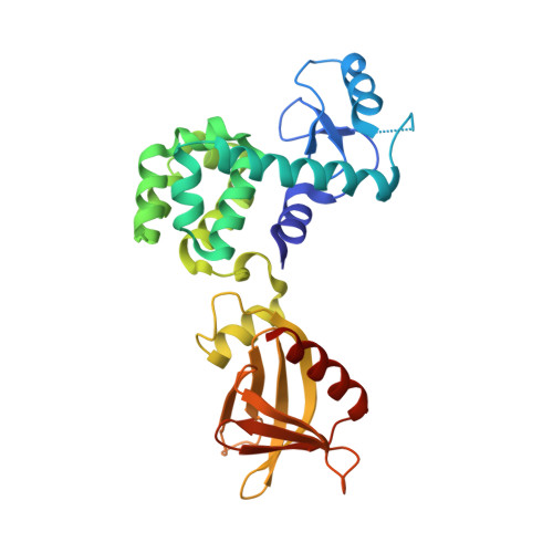
    Membrane contact sites (MCSs) in eukaryotic cells are hotspots for lipid exchange, which is essential for many biological functions, including regulation of membrane properties and protein trafficking. Lipid transfer proteins anchored at membrane contact sites (LAMs) contain sterol-specific lipid transfer domains [StARkin domain (SD)] and multiple targeting modules to specific membrane organelles. Elucidating the structural mechanisms of targeting and ligand recognition by LAMs is important for understanding the interorganelle communication and exchange at MCSs. Here, we determined the crystal structures of the yeast Lam6 pleckstrin homology (PH)-like domain and the SDs of Lam2 and Lam4 in the apo form and in complex with ergosterol. The Lam6 PH-like domain displays a unique PH domain fold with a conserved N-terminal α-helix. The Lam6 PH-like domain lacks the basic surface for phosphoinositide binding, but contains hydrophobic patches on its surface, which are critical for targeting to endoplasmic reticulum (ER)-mitochondrial contacts. Structures of the LAM SDs display a helix-grip fold with a hydrophobic cavity and a flexible Ω1-loop as a lid. Ergosterol is bound to the pocket in a head-down orientation, with its hydrophobic acyl group located in the tunnel entrance. The Ω1-loop in an open conformation is essential for ergosterol binding by direct hydrophobic interaction. Structural comparison suggested that the sterol binding mode of the Lam2 SD2 is likely conserved among the sterol transfer proteins of the StARkin superfamily. Structural models of full-length Lam2 correlated with the sterol transport function at the membrane contact sites.


  • Organizational Affiliation

    College of Pharmacy, Chonnam National University, Bukgu, Gwangju, 61186, Republic of Korea.


Macromolecules
Find similar proteins by:  (by identity cutoff)  |  3D Structure
Entity ID: 1
MoleculeChains Sequence LengthOrganismDetailsImage
Endolysin/Membrane-anchored lipid-binding protein LAM6 fusion protein276Tequatrovirus T4Saccharomyces cerevisiae S288C
This entity is chimeric
Mutation(s): 5 
EC: 3.2.1.17
Membrane Entity: Yes 
UniProt
Find proteins for P00720 (Enterobacteria phage T4)
Explore P00720 
Go to UniProtKB:  P00720
Find proteins for Q08001 (Saccharomyces cerevisiae (strain ATCC 204508 / S288c))
Explore Q08001 
Go to UniProtKB:  Q08001
Entity Groups  
Sequence Clusters30% Identity50% Identity70% Identity90% Identity95% Identity100% Identity
UniProt GroupsP00720Q08001
Sequence Annotations
Expand
  • Reference Sequence
Small Molecules
Ligands 1 Unique
IDChains Name / Formula / InChI Key2D Diagram3D Interactions
2PE
Query on 2PE

Download Ideal Coordinates CCD File 
B [auth A]NONAETHYLENE GLYCOL
C18 H38 O10
YZUUTMGDONTGTN-UHFFFAOYSA-N
Experimental Data & Validation

Experimental Data

  • Method: X-RAY DIFFRACTION
  • Resolution: 2.40 Å
  • R-Value Free: 0.266 
  • R-Value Work: 0.218 
  • R-Value Observed: 0.221 
  • Space Group: P 21 21 21
Unit Cell:
Length ( Å )Angle ( ˚ )
a = 59.543α = 90
b = 60.103β = 90
c = 105.6γ = 90
Software Package:
Software NamePurpose
PHENIXrefinement
HKL-2000data reduction
HKL-2000data scaling
PHASERphasing

Structure Validation

View Full Validation Report


Entry History & Funding Information

Deposition Data


Funding OrganizationLocationGrant Number
National Research Foundation (Korea)Korea, Republic OfNRF-2017R1A2B4004914

Revision History  (Full details and data files)

  • Version 1.0: 2018-01-31
    Type: Initial release
  • Version 1.1: 2018-02-14
    Changes: Database references
  • Version 1.2: 2023-11-22
    Changes: Data collection, Database references, Refinement description